Copyright
©2014 Baishideng Publishing Group Co.
World J Gastroenterol. Mar 28, 2014; 20(12): 3383-3387
Published online Mar 28, 2014. doi: 10.3748/wjg.v20.i12.3383
Published online Mar 28, 2014. doi: 10.3748/wjg.v20.i12.3383
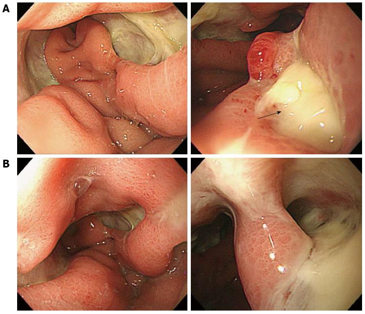
Figure 3 Esophagogastroduodenoscopy.
A: A large ulceration was detected (left), and perforation was suspected (right, black arrow), postoperative day 23; B: Submucosal dissection was suspected (left). Magnified image of the area of suspected dissection (right).
- Citation: Min SY, Kim YH, Park WS. Acute phlegmonous gastritis complicated by delayed perforation. World J Gastroenterol 2014; 20(12): 3383-3387
- URL: https://www.wjgnet.com/1007-9327/full/v20/i12/3383.htm
- DOI: https://dx.doi.org/10.3748/wjg.v20.i12.3383


