Copyright
©2014 Baishideng Publishing Group Co.
World J Gastroenterol. Mar 21, 2014; 20(11): 2810-2824
Published online Mar 21, 2014. doi: 10.3748/wjg.v20.i11.2810
Published online Mar 21, 2014. doi: 10.3748/wjg.v20.i11.2810
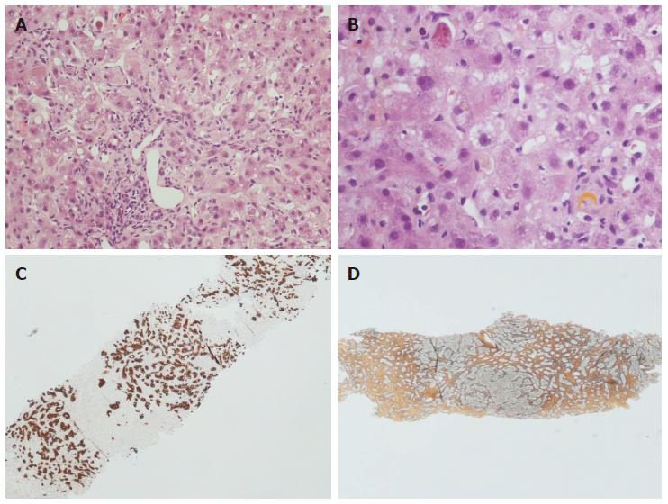
Figure 2 Histopathological appearance of fibrosing cholestatic recurrent hepatitis C.
Hematoxylin-eosin stain × 10 (A) and × 40 (B) magnification: note the lobular architectural disarray, the portal tract fibrosis and distortion, the lobular necrosis with Councilman bodies and the cholestasis with hepatocellular feathery degeneration and ballooning. The immunohistochemistry for keratin 19 (C) highlights the prominent ductular reaction, while the reticulin stain (D) indicates advanced fibrosis.
- Citation: Vasuri F, Malvi D, Gruppioni E, Grigioni WF, D’Errico-Grigioni A. Histopathological evaluation of recurrent hepatitis C after liver transplantation: A review. World J Gastroenterol 2014; 20(11): 2810-2824
- URL: https://www.wjgnet.com/1007-9327/full/v20/i11/2810.htm
- DOI: https://dx.doi.org/10.3748/wjg.v20.i11.2810


