Copyright
©2014 Baishideng Publishing Group Co.
World J Gastroenterol. Mar 21, 2014; 20(11): 2810-2824
Published online Mar 21, 2014. doi: 10.3748/wjg.v20.i11.2810
Published online Mar 21, 2014. doi: 10.3748/wjg.v20.i11.2810
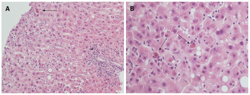
Figure 1 Typical histopathological appearance of acute recurrent hepatitis C.
A: Lobular architectural disarray, lobular necrosis with lymphocytic sinusoidal infiltrate and visible Councilman bodies (black arrow) are evident, as well as a mild portal tract inflammation (arrowhead); hematoxylin-eosin stain, × 20 magnification; B: Detail of the same case at × 40 magnification: note the high number of Councilman bodies in a single field (black arrows), and a minimal amount of macrovesicular steatosis.
- Citation: Vasuri F, Malvi D, Gruppioni E, Grigioni WF, D’Errico-Grigioni A. Histopathological evaluation of recurrent hepatitis C after liver transplantation: A review. World J Gastroenterol 2014; 20(11): 2810-2824
- URL: https://www.wjgnet.com/1007-9327/full/v20/i11/2810.htm
- DOI: https://dx.doi.org/10.3748/wjg.v20.i11.2810


