Copyright
©2014 Baishideng Publishing Group Co.
World J Gastroenterol. Mar 14, 2014; 20(10): 2586-2594
Published online Mar 14, 2014. doi: 10.3748/wjg.v20.i10.2586
Published online Mar 14, 2014. doi: 10.3748/wjg.v20.i10.2586
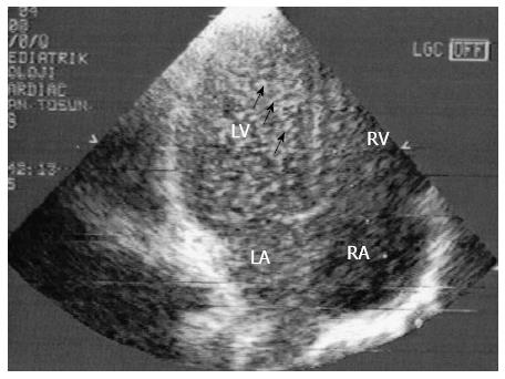
Figure 3 “Bubble” or contrast echocardiogram (apical four chamber view).
A stream of microbubbles filling the left atrium following systemic venous injection is shown.
- Citation: Tumgor G. Cirrhosis and hepatopulmonary syndrome. World J Gastroenterol 2014; 20(10): 2586-2594
- URL: https://www.wjgnet.com/1007-9327/full/v20/i10/2586.htm
- DOI: https://dx.doi.org/10.3748/wjg.v20.i10.2586


