Copyright
©2014 Baishideng Publishing Group Co.
World J Gastroenterol. Jan 7, 2014; 20(1): 319-322
Published online Jan 7, 2014. doi: 10.3748/wjg.v20.i1.319
Published online Jan 7, 2014. doi: 10.3748/wjg.v20.i1.319
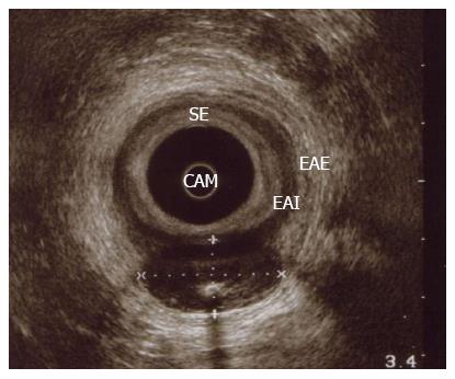
Figure 1 Endoanal ultrasound.
Posterior hipoecoid nodule, well circumscribed, between internal and external anal sphincter, with posterior enhancement and central calcification. CAM: Middle anal canal; EAI: Internal anal sphincter; EAE: External anal sphincter; SE: Subepithelium.
- Citation: Carvalho N, Albergaria D, Lebre R, Giria J, Fernandes V, Vidal H, Brito MJ. Anal canal gastrointestinal stromal tumors: Case report and literature review. World J Gastroenterol 2014; 20(1): 319-322
- URL: https://www.wjgnet.com/1007-9327/full/v20/i1/319.htm
- DOI: https://dx.doi.org/10.3748/wjg.v20.i1.319


