Copyright
©2014 Baishideng Publishing Group Co.
World J Gastroenterol. Jan 7, 2014; 20(1): 242-249
Published online Jan 7, 2014. doi: 10.3748/wjg.v20.i1.242
Published online Jan 7, 2014. doi: 10.3748/wjg.v20.i1.242
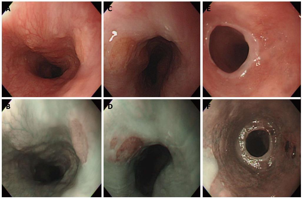
Figure 1 Endoscopic images of cervical esophageal heterotopic gastric mucosa.
A: Conventional imaging (CI) image of a typical heterotopic gastric mucosa (HGM) patch; B: Narrow-band imaging (NBI) appears to increase contrast and enhance the mucosal details between the columnar and squamous epithelia; C: Single HGM was found by CI image; D: In NBI contrast, additional small HGM was noted superior to the medium HGM; E: CI image in a patient with circumferential HGM and esophageal stenosis; F: With NBI, the columnar mucosa appears dark brown, and the squamous epithelia, light green. This sharp contrast of coloration aids in the detection of circumferential lesions.
- Citation: Cheng CL, Lin CH, Liu NJ, Tang JH, Kuo YL, Tsui YN. Endoscopic diagnosis of cervical esophageal heterotopic gastric mucosa with conventional and narrow-band images. World J Gastroenterol 2014; 20(1): 242-249
- URL: https://www.wjgnet.com/1007-9327/full/v20/i1/242.htm
- DOI: https://dx.doi.org/10.3748/wjg.v20.i1.242


