Published online Jan 7, 2014. doi: 10.3748/wjg.v20.i1.193
Revised: October 17, 2013
Accepted: November 2, 2013
Published online: January 7, 2014
Processing time: 227 Days and 19 Hours
AIM: To investigate the effects of diallyl trisulfide (DATS), a garlic-derived organosulfur compound, in pancreatic cancer cells.
METHODS: Human pancreatic cancer cells with wild-type p53 gene (Capan-2) and normal pancreatic epithelial cells (H6C7) were cultured in RPMI1640. DATS was prepared at a concentration of 100 μmol/L. Cell viability was determined via the methyl thiazolyl tetrazolium assay. Apoptotic cells were detected by TUNEL assay. Cell cycle analysis was performed using flow cytometry. Protein expression was determined by Western blot. Bax and Bcl-2 expression was detected by immunofluorescence. Apoptosis genes and cell cycle were assessed by quantitative real-time polymerase chain reaction.
RESULTS: DATS suppressed the viability of cultured human pancreatic cancer cells (Capan-2) by increasing the proportion of cells in the G2/M phase and induced apoptotic cell death. Western blot analysis indicated that DATS enhanced the expression of Fas, p21, p53 and cyclin B1, but downregulated the expression of Akt, cyclin D1, MDM2 and Bcl-2. DATS induced cell cycle inhibition which was correlated with elevated levels of cyclin B1 and p21, and reduced levels of cyclin D1 in Capan-2 cells and H6C7 cells. DATS-induced apoptosis was markedly elevated in Capan-2 cells compared with H6C7 cells, and this was correlated with elevated levels of cyclin B1 and p53, and reduced levels of Bcl-2. DATS-induced apoptosis was correlated with down-regulation of Bcl-2, Akt and cyclin D1 protein levels, and up-regulation of Bax, Fas, p53 and cyclin B protein levels in Capan-2 cells.
CONCLUSION: DATS induces apoptosis of pancreatic cancer cells (Capan-2) and non-tumorigenic pancreatic ductal epithelial cells (H6C7).
Core tip: We investigated the effect of diallyl trisulfide (DATS), a promising cancer chemopreventive constituent of processed garlic, in human pancreatic cancer cells (Capan-2) and non-tumorigenic pancreatic ductal epithelial cells (H6C7), and explored the possible mechanism involved. We found that DATS can induce apoptosis in both cell lines. We also investigated the role of p53, Bcl-2 and the cell cycle in apoptosis of Capan-2 and H6C7 cells.
- Citation: Ma HB, Huang S, Yin XR, Zhang Y, Di ZL. Apoptotic pathway induced by diallyl trisulfide in pancreatic cancer cells. World J Gastroenterol 2014; 20(1): 193-203
- URL: https://www.wjgnet.com/1007-9327/full/v20/i1/193.htm
- DOI: https://dx.doi.org/10.3748/wjg.v20.i1.193
The treatment of patients with pancreatic cancer mainly depends on surgery, radiation therapy, chemotherapy or combined therapeutic methods. Due to aggressive tumor biology and lack of specific symptoms during the early stages, pancreatic cancer frequently presents as an incurable disease with approximately two-thirds of patients showing radiographically detectable metastasis at the time of diagnosis. Pancreatic ductal adenocarcinomas carry a dismal prognosis, with fewer than 5% of patients surviving 5 years or more[1].
For patients without metastasis, the only potentially curative treatment is surgery. However, even among the 10% of patients with resectable disease, only about 20% survive for 5 years or more, indicating a need for good adjuvant therapies. Approximately 40% of patients diagnosed have locally advanced, unresectable, non-metastatic disease known as locally advanced pancreatic cancer. These patients are treated with chemoradiation therapy in the United States[2]. However, advanced pancreatic cancer is often resistant to conventional chemotherapy or radiotherapy[3].
The dismal prognosis of pancreatic cancer is further accentuated by its poor response to chemotherapy and to radiation therapy[4]. Thus, therapies are needed to increase the efficiency of radiation and chemotherapy. The discovery of new therapeutic agents and approaches in patients with pancreatic cancer is of paramount importance.
Genetic changes in apoptotic regulatory mechanisms may result in an increase in cell numbers, as well as the preservation of genetically altered cells, which initiate the process of tumorigenesis[5]. Disordered apoptosis and abnormal proliferation have been linked to the development of malignancy and treatment resistance[6]. Apoptosis, also termed programmed cell death, occurs via extrinsic or intrinsic signal transduction pathways[7]. Therefore, further understanding of the molecular mechanisms of apoptosis and the relationship between pancreatic cancer chemoresistance and disordered apoptosis and abnormal proliferation is needed. Furthermore, apoptosis contributes to cell death in tumors treated with various anticancer agents. Chemotherapy, radiation therapy and immunotherapy all rely heavily on the induction of apoptosis to kill pancreatic cancer cells.
Many recent studies have revealed that certain garlic-derived organosulfur compounds can suppress the proliferation of cultured cancer cells by causing apoptosis and/or cell cycle arrest[8-10].
Garlic (Allium sativum) is a common plant used mainly as food and has recently been reported to have medicinal properties[11]. Garlic-derived organosulfur compounds including diallyl sulfide, diallyl disulfide (DADS) and/or diallyl trisulfide (DATS), which are major components of garlic, may be associated with a reduced risk of certain cancers[12].
These compounds are known to inhibit cell proliferation, cause cell cycle arrest and increase apoptosis in various cancer cell lines[13]. However, the cellular and molecular mechanisms underlying these activities have not yet been completely elucidated.
In the present study, we investigated the effects of the garlic constituent, DATS, in human pancreatic cancer cells (Capan-2) and non-tumorigenic pancreatic ductal epithelial cells (H6C7).
The human pancreatic cancer cell line Capan-2, with wild-type p53 gene, and the normal pancreatic epithelial cell line H6C7 were obtained from ATCC (United States) and cultured in RPMI1640 (Gibco, CA, United States) containing 10% heat-inactivated fetal bovine serum (FBS), benzylpenicillin (100 KU/L), streptomycin (100 mg/L), transferrin (0.001 mg/mL), EGF (20 ng/μL) and hydrocortisone (500 ng/mL) at 37 °C in a humidified incubator containing 5% CO2 in air (Gibco). The cells were cultured in plastic culture dishes with a non-adhesive surface and digested with 0.25% trypsin for subculture.
DATS (99% purity) was purchased from Chia-Tai Tianqing Pharmaceutical Co., Ltd. (China). DATS was prepared at a concentration of 100 μmol/L and dissolved in suitable medium.
Cell viability was detected using the methyl thiazolyl tetrazolium (MTT) (Sigma, St. Louis, MO, United States) assay. Briefly, cells in the logarithmic growth phase at a density of 1 × 10⁴ cells/well in 100 μL of medium were seeded in a 96-well plate and cultured for 24 h prior to drug treatment. The medium was then changed to medium containing (100 μmol/L) DATS. After 24 h, 10 μL of 5 mg/mL MTT reagent was added to each well and incubated for 4 h. After incubation, 100 μL of detergent reagent was added to each well to dissolve the formazan crystals. The absorbance of the solution was determined at 570 nm (A570 nm) and the cell inhibition ratio was calculated as follows: cell inhibition ratio = [(A570 nm value of the control group - A570 nm value of the experimental group)/A570 nm value of the control group] × 100%. Cells treated with irradiation only were used as controls. Each assay was performed in triplicate and the standard deviation was determined.
Apoptotic cells were determined by TUNEL assay using the In Situ Cell Death Detection kit (Roche, Mannheim, Germany) according to the manufacturer’s instructions. Cells were cultured on 4-chamber slides (VWR, United States) at a density of 2 × 10⁴ cells/chamber. After treatment with 100 μmol/L of DATS, the cells were washed with PBS and fixed with 4% paraformaldehyde (Sigma-Aldrich Corp., United States) for 20 min at 4 °C. The fixed cells were incubated with digoxigenin-conjugated dUTP in a terminal deoxynucleotide transferase recombinant (rTdT)-catalyzed reaction and nucleotide mixture for 1 h at 37 °C in a humidified atmosphere, and then immersed in stop/wash buffer for 15 min at room temperature. Cells were then washed with PBS to remove unincorporated fluorescein-12-dUTP. After washing with PBS, cells were incubated with 1 mg/mL DAPI (Sigma) solution for 30 min at 37 °C in the dark. Apoptotic cells were photographed under a fluorescence microscope (Nikon Inc., Tokyo, Japan).
Cells were seeded at a density of 3-5 × 10⁵/plate and incubated for 24 h before irradiation. The medium was changed to medium containing DATS (100 μmol/L) 30 min before irradiation. Cells were harvested by trypsinization after 24 h. The cells were washed with PBS and then fixed with ice-cold 70% ethanol while vortexing. Finally, the cells were washed and resuspended in PBS containing 5 μg/mL RNase A (Sigma) and 50 μg/mL propidium iodide (Sigma) for analysis. Cell cycle analysis was performed using a FACScan Flow Cytometer (Becton Dickson, United States) according to the manufacturer’s protocol.
The samples were normalized to monitor protein concentrations and the loading dye (50 mmol/L Tris-Cl pH 6.8, 100 mmol/L β-mercaptoethanol, 2% sodium dodecyl sulfate (SDS), 0.1% bromophenol blue, 10% glycerol) was added. The proteins were separated by 12.5% sodium dodecyl sulfate-polyacrylamide gel electrophoresis and transferred to a polyvinylidene fluoride membrane (Millipore, United States). After transferring and blocking, the membrane was incubated with an appropriate primary antibody (Santa Cruz Biotechnology) at a 1:1000 dilution in PBS-T containing 5% milk overnight at 4 °C. Before the transfer, the membrane was activated by incubation in methanol for 30 s, water for 2 min and then transfer buffer (48 mmol/L Tris base, 20% methanol, 0.04% SDS, 30 mmol/L glycine) until ready for transfer. A semi-dry transfer apparatus (Owl Separation Systems, Portsmouth, NH, United States) was used to transfer the proteins onto the membrane. The membrane was quickly washed three times, then two 5 min washes and incubated with the appropriate secondary antibody conjugated to horseradish peroxidase (goat anti-mouse antibody, Santa Cruz Biotechnology).
For Western blot analysis, mouse anti-human cyclin D1, p21, cyclin B1, caspase-3, caspase-8, Bcl-2, Bax, and Fas monoclonal antibodies (1:1000 for all) were used. Anti-glyceraldehyde phosphate dehydrogenase (GAPDH) and anti-β-actin antibodies (1:1000; Santa Cruz Biotechnology) were used to detect internal controls. All experiments were performed in triplicate.
Capan-2 cells in 5% CS and 1% penicillin streptomycin-containing Dulbecco’s modification of Eagle’s medium Dulbecco (DMEM) were cultured in two-well chambers and then mounted on glass slides with plastic covers. At 70% confluence, the cells were switched to fresh 5% CS and 1% penicillin streptomycin-containing DMEM medium supplemented with or without 100 μmol/L DATS. After fixation and washing, the cells were incubated with the primary antibody Bp53-12 (Santa Cruz Biotechnology). Following overnight incubation, the cells were incubated with the secondary antibody (donkey anti-mouse IgG) (Santa Cruz Biotechnology). Finally, the cells were visualized and photographed at × 400 magnification using an immunofluorescence microscope (Nikon Inc., Tokyo, Japan). The images were merged using the advanced SPOT software (Diagnostic Instruments Inc., Sterling Heights, MI, United States). For the detection of Bax, the primary antibody which was diluted at 1:400 in 2% chicken serum/PBS was added. Cells were then washed three times with PBS and incubated with Texas Red-conjugated secondary antibody (Santa Cruz Biotechnology).
Capan-2 cells were cultured in 5% CS and 1% penicillin streptomycin-containing DMEM to 70% confluence and then switched to new culture medium supplemented with 100 μmol/L DATS. After 48 h, RNA was extracted using the RNAeasy kit according to the manufacturer’s protocol. The extracted total RNA was subjected to cDNA synthesis using the Biorad iScriptTM Select cDNA Synthesis kit (Biorad, Hercules, CA, United States) containing random and oligo (dT) primer mix. The cDNA produced was used to specifically quantify the transcript of interest using RT-PCR. Quantitative RT-PCR was performed using SYBR Green chemistry (SYBR Green PCR Master Mix (Applied Biosystems, Foster City, CA, United States). The increase in fluorescence of the SYBR Green dye was monitored using a 7500 Real-Time PCR System (Applied Biosystems). The primers were: forward, 5’-ACC ACA GTC CAT GCC ATC AC-3’ and reverse, 5’-TCC ACC ACC CTG TTG CTG TA-3’ for the house-keeping gene GAPDH; forward, 5’-AAG CTG TGC ATC TAC ACC GA-3’ and reverse, 5’-CTT GAG CTT GTT CAC CAG GA-3’ for cyclin D1; forward, 5’-AAC ACC ATG GAC AGG GAG AG -3’ and reverse, 5’-CCC AGC AGC TTC AGG TAC TC -3’ for Akt; forward, 5’-GGC TCA GCA TGG TCG CTT-3’ and reverse, 5’-CTC CCG CCA GCT GTC ATT-3’ for Fas; forward, 5’-TTG AGT TCG GTG GGG TCA TG-3’ and reverse, 5’-GAT CCA GGT GTG CAG ATG CC-3’ for BcL-2; forward, 5’-CTG AGC TGA CCT TGG AGC-3’ and reverse, 5’-GAC TCC AGC CAC AAA GAT G-3’ for Bax. The SyBr green dye intercalates with the double-stranded cDNA formed during PCR, allowing easy quantification of cDNA to indirectly quantify the original RNA transcripts. Thermal cycling conditions were designed as follows: reverse transcription at 50 °C for 10 min followed by an initial denaturation at 95 °C for 5 min and 35 cycles of 95 °C for 15 s and 60 °C for 30 s. The comparative CT method (ΔΔCt) was used to study the relative quantification of gene expression. Each evaluation was performed in triplicate in three independent experiments.
All the assays described above were repeated more than once. The quantification assays were performed at least twice and data were calculated and presented as mean ± SD. Data were either analyzed by unpaired Student’s t test or one-way ANOVA. Differences were considered significant at P < 0.05.
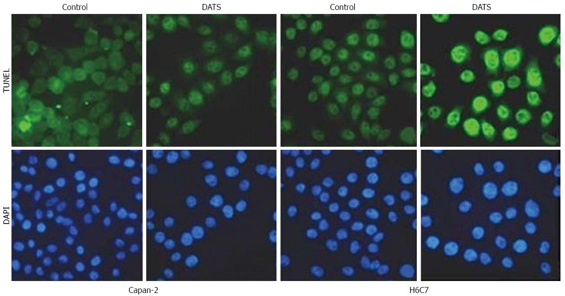
In Capan-2 cells and H6C7 cells, TUNEL assay were performed to ascertain the induction of apoptosis by 100 μmol/L DATS. Fewer TUNEL-positive cells were found in H6C7 cells that in Capan-2 cells after treatment with 100 μmol/L of DATS (Figure 1).
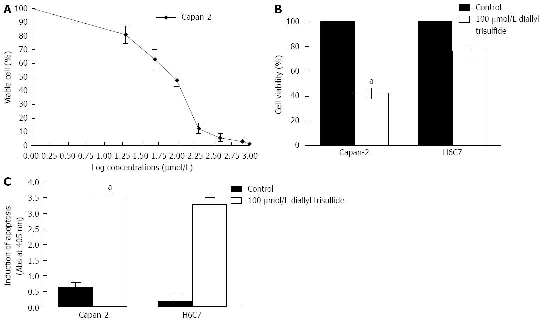
The effect of DATS on cell viability and cell apoptosis induction in Capan-2 cells was examined by MTT assay. A dose-response curve was constructed from which we chose 100 μmol/L for subsequent experiments (Figure 2A). The analysis revealed that 100 μmol/L of DATS decreased the viability of both Capan-2 cells (55%) and H6C7 cells (30%) compared with untreated control cells (P < 0.05) (Figure 2B). ELISA indicated that 100 μmol/L of DATS induced apoptosis of Capan-2 cells (about an 8-fold increase) compared with controls. In addition, the viability of H6C7 cells was significantly decreased by about 5 folds (P < 0.05) (Figure 2C).
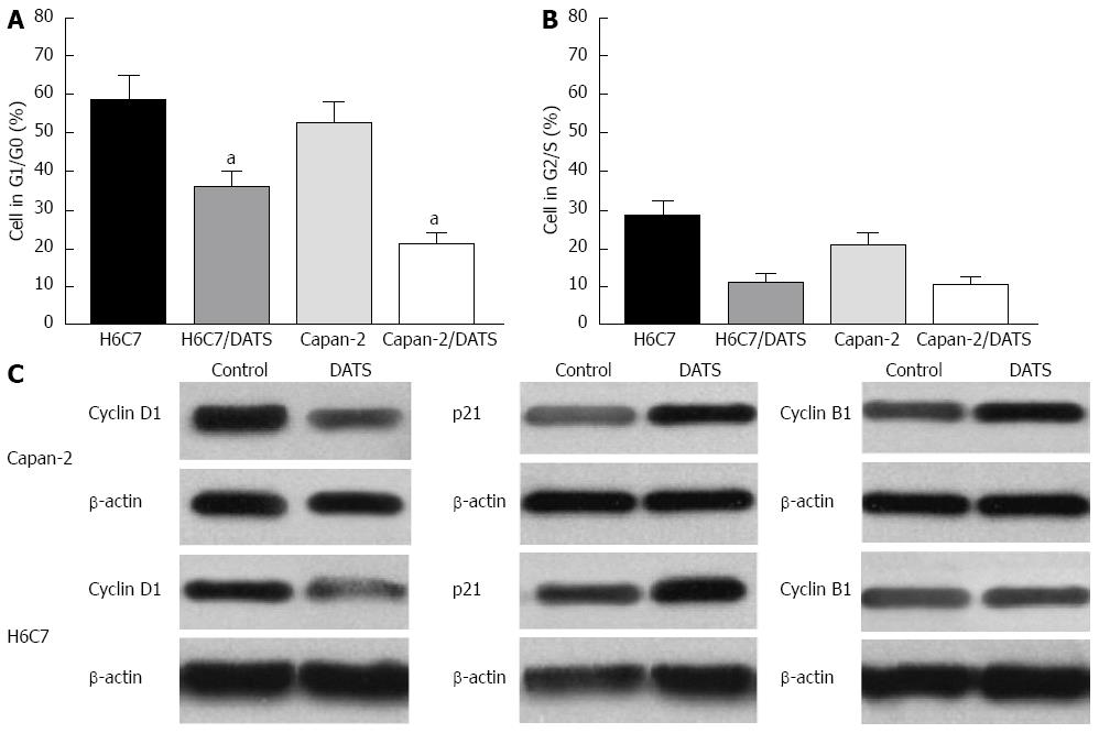
Flow cytometry was performed to study the effects of DATS on cell cycle progression. Treatment of both cell lines was carried out in three independent experiments and is represented in a histogram. The percentages of cells in G1/G0 and G2/S were determined after treatment with 100 μmol/L DATS for 24 h. Both Capan-2 and H6C7 cells treated with DATS and harvested after 24 h showed an decrease in the percentage of G1/G0 cells compared with control cells (P < 0.05), and the reduced value in Capan-2 cells was about 35% (Figure 3A). No significant difference was found in the percentage of cells in the G2/S phase in both Capan-2 and H6C7 cells compared with control cells (Figure 3B). The expression of cyclin D1, p21 and cyclin B1 was determined by Western blot in Capan-2 and H6C7 cells. The results showed that DATS decreased the level of cyclin D1 and increased the levels of cyclin B1 and p21 in both Capan-2 and H6C7 cells (Figure 3C).
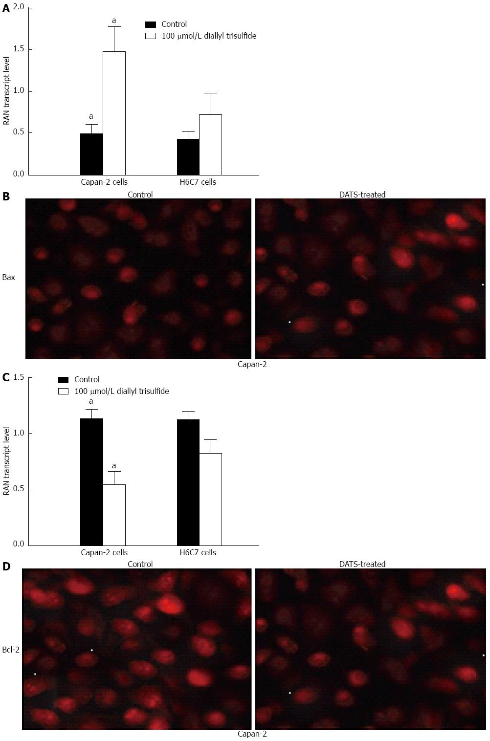
Capan-2 cells were treated with 100 μmol/L DATS for 24 h. Using RT-PCR and Western blot analyses, it was found that DATS significantly increased Bax expression at the mRNA level by 3 folds and decreased Bcl-2 expression at the mRNA level by 0.5 folds as indicated in Figure 4A and C (P < 0.05). These results were further confirmed by immunofluorescence (Figure 4B and D).
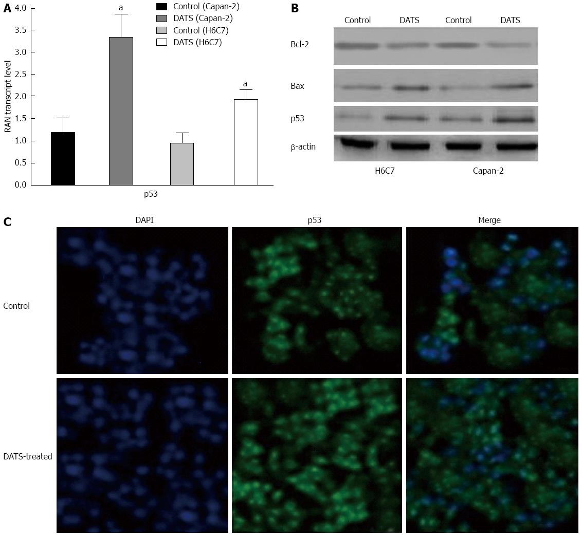
The p53 expression was up-regulated in Capan-2 cells compared with that in H6C7 cells after treatment with 100 μmol/L DATS. Bax expression was also elevated in Capan-2 cells by approximately 3 folds compared with the untreated control group (P < 0.05); however, Bcl-2 expression was reduced in Capan-2 cells (Figure 5A and B). Immunofluorescence showed that treatment with 100 μmol/L DATS induced p53 translocation from the cytoplasm (indicated in green) to the nucleus (indicated in blue) compared with the untreated control group (Figure 5C).
Akt inhibits molecular events that precede cytochrome C release, while Bcl-2 prevents its initial release. Treatment with 100 μmol/L DATS for 24 h significantly decreased the expression level of Akt by 40% compared with the control group (Figure 6B, P < 0.055). FAS expression level was increased after DATS treatment for 24 h by 4 folds compared with the control group (Figure 6, P < 0.05). Figure 6B also shows a 2-fold increase in the expression level of cyclin D1 (P < 0.05) and a 50% decrease in Bcl-2 expression level in response to 100 μmol/L DATS treatment for 24 h (P < 0.05) in Capan-2 cells. The results of RT-PCR also confirmed the above findings with a decrease in MDM2 level and an increase in p53 level as previously described (Figure 6A).
Allium vegetables have been used in traditional medicine for centuries[14]. Recent scientific investigations have shown that allium vegetables and their constituents, garlic-derived organosulfur compounds (OSCs), including diallyl sulfide, diallyl disulfide and DATS, reduce the risk of cardiovascular disease and diabetes, stimulate the immune system, protect against infections, and have anti-aging as well as anti-cancer effects[13]. The anticancer effects of allium vegetables, especially garlic, are supported by epidemiological data[15-17]. It has been proved that garlic-derived OSCs, including diallyl sulfide, diallyl disulfide and DATS, show significant protection against different types of malignancies in animal models[18-24].
Numerous publications indicate that the suppression of cancer cell growth by OSCs correlates with apoptosis induction. Elucidation of the mechanism(s) of apoptosis induction by OSCs has been the topic of intense research in the last few years. Most studies indicate the involvement of Bcl-2 family proteins and p53 in the regulation of OSC-mediated apoptosis[9,24-27]. Our data showed that DATS treatment could induce apoptosis of Capan-2 cells via activation of p53. p53 is a tumor suppressor, is mutated in 50% of human cancers and regulates cell growth and sensitivity to irradiation and multiple anticancer agents[28-30]. Functional p53 can downregulate Bcl-2, which allows cells to survive a variety of fatal cellular events and protects cells from apoptosis[31]. p53 can also induce p21, and an increased level of p21 can in turn decrease the activity of cyclin-dependent kinases (CDKs), resulting in growth arrest[32-34]. Our results indicated that DATS increased the expression and translocation of p53 from the cytoplasm to the nucleus in Capan-2 cells following exposure to DATS, which was correlated with increased induction of apoptosis.
The tumor suppressor p53 is a key regulator of apoptosis and has pro-apoptotic activity. Under stress conditions, p53 is stabilized and acts as a transcription factor which may increase the expression of pro-apoptotic target genes, such as Puma, Noxa, Bax and Bid[35]. On the other hand, cytoplasmic p53 interacts with Bcl-2 family members, Bcl-2 or Bcl-xL, which results in activation and translocation of Bax and Bid to the mitochondrial outer membrane. Moreover, p53 also translocates to the mitochondria to activate the mitochondrial apoptosis pathway[35-37]. It was reported that DATS-induced apoptosis was attributed to the induction of Bax, downregulation of Bcl-xL and activation of caspase 3[8,38]. In addition, activated Akt can phosphorylate several apoptosis-regulating proteins including pro-apoptotic Bcl-2 family member, BAD[39,40]. BAD promotes cell death by interacting with anti-apoptotic Bcl-2 members such as Bcl-xL, which allows the multidomain pro-apoptotic Bcl-2 family members, Bax and Bak, to aggregate and cause release of apoptogenic molecules (e.g., cytochrome C) from mitochondria to the cytosol, culminating in caspase activation and cell death[41]. Our results support the conclusion that a significant decrease in the levels of survival signals is generated by both Bcl-2 and Akt (Figure 6).
It is well documented that inappropriate expression of cell-cycle regulatory proteins can contribute to human tumorigenesis[42]. Numerous studies have reported the relationship between carcinogenesis and cell cycle-related genes[43-47]. In experimental animals, DATS was capable of suppressing the growth of cancer cells in culture[43]. The DATS-mediated inhibition of cancer cell proliferation correlated with cell cycle arrest and apoptosis induction[42]. The mechanism of G2 phase arrest remains elusive. It was demonstrated that the DATS-mediated G2/M phase cell cycle arrest in the human prostate cancer cell line DU145 was transient and caused by differential kinetics of nuclear localization of CDK1 and cyclin B1[44]. The DATS-treated prostate cancer cells were arrested in G2 as well as the prometaphase[45]. This DATS-mediated prometaphase arrest was caused by checkpoint kinase 1-mediated inactivation of anaphase promoting complex/cyclosome characterized by accumulation of its substrates (e.g., securin)[42,46]. A fraction of cells arrested in prometaphase following treatment with DATS are ultimately driven to apoptotic cell death[46]. However, the mechanism underlying DATS-mediated G2 arrest is unclear.
The present study aimed to introduce DATS as an inducer of apoptosis in human pancreatic cancer cells (Capan-2) via a decrease in the percentage of cells in G2/M phase. DATS also increased the sensitivity of Capan-2 cells to radiation as indicated by the clonogenic survival assay and confirmed by flow cytometric analysis. Our data indicated that DATS increased the level of cyclin B1 in both Capan-2 and H6C7 cells, which was correlated with G2/M arrest, shown by a decrease in the percentage of cells in G2/M phase compared with the control group. Consistent with our data, it was recently reported that DATS suppresses the viability of cultured human lung cancer cells and normal human bronchial epithelial cells (BEAS-2B) by causing G2-M phase arrest and apoptotic cell death[47]. It is possible that the G2-M arrest is a priming mechanism for initiation of the cell death process in Capan-2 and H6C7 cells. A checkpoint kinase 1-dependent mechanism for DATS-induced mitotic arrest in human prostate cancer cells was previously reported[45]. Among the possible mechanisms for increased sensitivity to apoptosis in Capan-2 cells, overexpression of cyclin B1 may decrease the percentage of cells in G2/M phase. Our results also showed that elevated levels of cyclin B1 protein in Capan-2 and H6C7 cells due to DATS treatment increased apoptosis (Figure 4).
Cell cycle progression and cell division are driven by the sequential activation of a group of serine-threonine kinases called cyclin-dependent kinases (Cdks). Multiple Cdks control the cell cycle in mammals and have long been considered essential for normal proliferation, development and homeostasis[48]. Cyclin B1 is the regulatory subunit of the M-phase promoting factor, and correct regulation of cyclin B1 is essential for the initiation of mitosis. The deregulation of cyclin B1 is involved in neoplastic transformation, and the suppression of cyclin B1 could be an attractive strategy in antiproliferative therapy[49]. Our results also showed that elevated levels of cyclin B1 protein in Capan-2 and H6C7 cells due to DATS treatment increased apoptosis (Figure 4). Conversely, downregulation of cyclin B1 consequently reduced the activity of CDK1/cyclin B1 and blocked the aggressive proliferation of tumor cells[50].
There may be distinct differences in cyclin B1 signaling between cancer cell types, as a decrease in cyclin B1 protein signal pathways leads to apoptosis of cervical carcinoma cells, but not prostate cancer cells. Therefore, gene therapy strategies that reduce cyclin B1 protein are not likely to be effective in all types of cancer.
Cyclin D1 accumulates and activates its cognate CDK (CDK4/6) in response to mitogenic growth factors in early to mid G1 phase[51]. Perturbations in this step, which result in reduced growth factor requirements for cyclin D1/CDK activation, will provide cells with a distinct growth advantage over their normal counterparts, and thus likely represents an early event in neoplasia. Our data showed an increase in the expression level of cyclin D1, which was correlated with the percentage decrease in cells in the G2/M phase and an increase in the induction of apoptosis.
The enhanced induction of apoptosis after ionizing radiation was related to the induction of p53 and p21 by cyclin D1[52]. In the present study, cyclin D1-associated overexpression of p53 may facilitate DATS-induced death of Capan-2 cells. The mechanism for DATS-induced apoptosis has been studied in various cellular systems, and it is unclear if the DATS-induced apoptosis signaling in Capan-2 cancer cells resembles that in other cellular systems. The results of the present study point to noticeable differences in apoptosis signaling between Capan-2 cells and other cancer cells.
In conclusion, the present study indicated that DATS treatment inhibited the growth of human pancreatic cancer cells (Capan-2) by inducing apoptosis. In addition, non-tumorigenic pancreatic ductal epithelial cells (H6C7) were significantly more resistant to DATS-mediated growth suppression and apoptosis induction compared with pancreatic cancer cells (Capan-2). Also, our data showed that DATS increased the expression of p53 and induced its translocation to the nucleus in Capan-2 cells. DATS also increased the expression of Bax as a downstream target of p53. We also provided evidence to indicate the involvement of Fas, cyclin B1, cyclin D, Akt and Bcl-2 as possible critical mediators of DATS-induced apoptosis signaling in Capan-2 cells.
Many recent studies have revealed that certain garlic-derived organosulfur compounds can suppress proliferation of cultured cancer cells by causing apoptosis and/or cell cycle arrest. Garlic (Allium sativum) is a common plant used mainly as food and has recently been reported to have medicinal properties. Garlic-derived organosulfur compounds including diallyl sulfide, diallyl disulfide (DADS) and/or diallyl trisulfide (DATS), which are major components of garlic, may be associated with a reduced risk of certain cancers. These compounds are known to inhibit cell proliferation, cause cell cycle arrest and increase apoptosis in various cancer cell lines. However, the cellular and molecular mechanisms underlying these activities have not yet been completely elucidated.
The dismal prognosis of pancreatic cancer is further accentuated by its poor response to chemotherapy and to radiation therapy. Thus, therapies are needed to increase the efficiency of radiation and chemotherapy. The discovery of new therapeutic agents and approaches in patients with pancreatic cancer is of paramount importance. DATS may be a novel strategy for the treatment of human pancreatic cancer through radio- or chemo-sensitization.
The present study indicates that DATS treatment inhibits the growth of human pancreatic cancer cells (Capan-2) by inducing apoptosis. In addition, non-tumorigenic pancreatic ductal epithelial cells (H6C7) are significantly more resistant to DATS-mediated growth suppression and apoptosis induction than Capan-2 cells. The data also showed that DATS increased the expression of p53 and induced its translocation to the nucleus in human Capan-2 cells. DATS also increased the expression of Bax as a downstream target of p53. This study provided evidence to indicate the involvement of Fas, cyclin B1, cyclin D, Akt and Bcl-2 as possible critical mediators of DATS-induced apoptosis signaling in Capan-2 cells.
This study demonstrates that DATS, a promising cancer chemopreventive constituent of processed garlic, induces apoptosis in human pancreatic cancer cells (Capan-2) and non-tumorigenic pancreatic ductal epithelial cells (H6C7). The results show, for the first time, that DATS administration might be a novel strategy for the treatment of human pancreatic cancer.
Garlic-derived organosulfur compounds including diallyl sulfide, DADS and/or DATS, which are major components of garlic, may be associated with a reduced risk of certain cancers.
In this report, the authors present data demonstrating a pattern of cell survival and gene expression changes suggesting that diallyl disulfide may provide therapeutic benefit against pancreatic ductal adenocarcinoma. The data are presented nicely and the findings merit publication.
P- Reviewer: Russ AJ S- Editor: Zhai HH L- Editor: Wang TQ E- Editor: Ma S
| 1. | Siegel R, Naishadham D, Jemal A. Cancer statistics, 2012. CA Cancer J Clin. 2012;62:10-29. [RCA] [PubMed] [DOI] [Full Text] [Cited by in Crossref: 8406] [Cited by in RCA: 8966] [Article Influence: 689.7] [Reference Citation Analysis (0)] |
| 2. | Deorukhkar A, Shentu S, Park HC, Diagaradjane P, Puduvalli V, Aggarwal B, Guha S, Krishnan S. Inhibition of radiation-induced DNA repair and prosurvival pathways contributes to vorinostat-mediated radiosensitization of pancreatic cancer cells. Pancreas. 2010;39:1277-1283. [RCA] [PubMed] [DOI] [Full Text] [Cited by in Crossref: 15] [Cited by in RCA: 15] [Article Influence: 1.0] [Reference Citation Analysis (0)] |
| 3. | Shen L, Kim SH, Chen CY. Sensitization of human pancreatic cancer cells harboring mutated K-ras to apoptosis. PLoS One. 2012;7:e40435. [RCA] [PubMed] [DOI] [Full Text] [Full Text (PDF)] [Cited by in Crossref: 10] [Cited by in RCA: 11] [Article Influence: 0.8] [Reference Citation Analysis (0)] |
| 4. | Farhana L, Dawson MI, Das JK, Murshed F, Xia Z, Hadden TJ, Hatfield J, Fontana JA. Adamantyl Retinoid-Related Molecules Induce Apoptosis in Pancreatic Cancer Cells by Inhibiting IGF-1R and Wnt/β-Catenin Pathways. J Oncol. 2012;2012:796729. [RCA] [PubMed] [DOI] [Full Text] [Full Text (PDF)] [Cited by in Crossref: 18] [Cited by in RCA: 22] [Article Influence: 1.7] [Reference Citation Analysis (0)] |
| 5. | Furth PA. Apoptosis and the development of breast cancer. Bowcock AM. Breast cancer: molecular genetics, pathogenesis and therapeutics. New Jersey: Humana Press 1999; 171-180. |
| 6. | Hanahan D, Weinberg RA. Hallmarks of cancer: the next generation. Cell. 2011;144:646-674. [RCA] [PubMed] [DOI] [Full Text] [Cited by in Crossref: 51728] [Cited by in RCA: 46852] [Article Influence: 3346.6] [Reference Citation Analysis (5)] |
| 7. | Qiao L, Wong BC. Targeting apoptosis as an approach for gastrointestinal cancer therapy. Drug Resist Updat. 2009;12:55-64. [RCA] [PubMed] [DOI] [Full Text] [Cited by in Crossref: 100] [Cited by in RCA: 106] [Article Influence: 6.6] [Reference Citation Analysis (0)] |
| 8. | Xiao D, Singh SV. Diallyl trisulfide, a constituent of processed garlic, inactivates Akt to trigger mitochondrial translocation of BAD and caspase-mediated apoptosis in human prostate cancer cells. Carcinogenesis. 2006;27:533-540. [PubMed] |
| 9. | Kim YA, Xiao D, Xiao H, Powolny AA, Lew KL, Reilly ML, Zeng Y, Wang Z, Singh SV. Mitochondria-mediated apoptosis by diallyl trisulfide in human prostate cancer cells is associated with generation of reactive oxygen species and regulated by Bax/Bak. Mol Cancer Ther. 2007;6:1599-1609. [PubMed] |
| 10. | Stan SD, Singh SV. Transcriptional repression and inhibition of nuclear translocation of androgen receptor by diallyl trisulfide in human prostate cancer cells. Clin Cancer Res. 2009;15:4895-4903. [RCA] [PubMed] [DOI] [Full Text] [Full Text (PDF)] [Cited by in Crossref: 32] [Cited by in RCA: 28] [Article Influence: 1.8] [Reference Citation Analysis (0)] |
| 11. | Gayathri R, Gunadharini DN, Arunkumar A, Senthilkumar K, Krishnamoorthy G, Banudevi S, Vignesh RC, Arunakaran J. Effects of diallyl disulfide (DADS) on expression of apoptosis associated proteins in androgen independent human prostate cancer cells (PC-3). Mol Cell Biochem. 2009;320:197-203. [RCA] [PubMed] [DOI] [Full Text] [Cited by in Crossref: 25] [Cited by in RCA: 27] [Article Influence: 1.6] [Reference Citation Analysis (0)] |
| 12. | Kaschula CH, Hunter R, Parker MI. Garlic-derived anticancer agents: structure and biological activity of ajoene. Biofactors. 2010;36:78-85. [RCA] [PubMed] [DOI] [Full Text] [Cited by in Crossref: 12] [Cited by in RCA: 27] [Article Influence: 1.8] [Reference Citation Analysis (0)] |
| 13. | Powolny AA, Singh SV. Multitargeted prevention and therapy of cancer by diallyl trisulfide and related Allium vegetable-derived organosulfur compounds. Cancer Lett. 2008;269:305-314. [RCA] [PubMed] [DOI] [Full Text] [Full Text (PDF)] [Cited by in Crossref: 212] [Cited by in RCA: 178] [Article Influence: 10.5] [Reference Citation Analysis (0)] |
| 15. | You WC, Blot WJ, Chang YS, Ershow A, Yang ZT, An Q, Henderson BE, Fraumeni JF, Wang TG. Allium vegetables and reduced risk of stomach cancer. J Natl Cancer Inst. 1989;81:162-164. [PubMed] |
| 16. | Zhou Y, Zhuang W, Hu W, Liu GJ, Wu TX, Wu XT. Consumption of large amounts of Allium vegetables reduces risk for gastric cancer in a meta-analysis. Gastroenterology. 2011;141:80-89. [RCA] [PubMed] [DOI] [Full Text] [Cited by in Crossref: 131] [Cited by in RCA: 111] [Article Influence: 7.9] [Reference Citation Analysis (0)] |
| 17. | Hsing AW, Chokkalingam AP, Gao YT, Madigan MP, Deng J, Gridley G, Fraumeni JF. Allium vegetables and risk of prostate cancer: a population-based study. J Natl Cancer Inst. 2002;94:1648-1651. [PubMed] |
| 18. | Wargovich MJ. Diallyl sulfide, a flavor component of garlic (Allium sativum), inhibits dimethylhydrazine-induced colon cancer. Carcinogenesis. 1987;8:487-489. [PubMed] |
| 19. | Wargovich MJ, Woods C, Eng VW, Stephens LC, Gray K. Chemoprevention of N-nitrosomethylbenzylamine-induced esophageal cancer in rats by the naturally occurring thioether, diallyl sulfide. Cancer Res. 1988;48:6872-6875. [PubMed] |
| 20. | Takahashi S, Hakoi K, Yada H, Hirose M, Ito N, Fukushima S. Enhancing effects of diallyl sulfide on hepatocarcinogenesis and inhibitory actions of the related diallyl disulfide on colon and renal carcinogenesis in rats. Carcinogenesis. 1992;13:1513-1518. [PubMed] |
| 21. | Reddy BS, Rao CV, Rivenson A, Kelloff G. Chemoprevention of colon carcinogenesis by organosulfur compounds. Cancer Res. 1993;53:3493-3498. [PubMed] |
| 22. | Schaffer EM, Liu JZ, Green J, Dangler CA, Milner JA. Garlic and associated allyl sulfur components inhibit N-methyl-N-nitrosourea induced rat mammary carcinogenesis. Cancer Lett. 1996;102:199-204. [PubMed] |
| 23. | Suzui N, Sugie S, Rahman KM, Ohnishi M, Yoshimi N, Wakabayashi K, Mori H. Inhibitory effects of diallyl disulfide or aspirin on 2-amino-1-methyl-6-phenylimidazo[4,5-b]pyridine-induced mammary carcinogenesis in rats. Jpn J Cancer Res. 1997;88:705-711. [PubMed] |
| 24. | Malki A, El-Saadani M, Sultan AS. Garlic constituent diallyl trisulfide induced apoptosis in MCF7 human breast cancer cells. Cancer Biol Ther. 2009;8:2175-2185. [PubMed] |
| 25. | Yang JS, Chen GW, Hsia TC, Ho HC, Ho CC, Lin MW, Lin SS, Yeh RD, Ip SW, Lu HF. Diallyl disulfide induces apoptosis in human colon cancer cell line (COLO 205) through the induction of reactive oxygen species, endoplasmic reticulum stress, caspases casade and mitochondrial-dependent pathways. Food Chem Toxicol. 2009;47:171-179. [RCA] [PubMed] [DOI] [Full Text] [Cited by in Crossref: 89] [Cited by in RCA: 92] [Article Influence: 5.4] [Reference Citation Analysis (0)] |
| 26. | Hong YS, Ham YA, Choi JH, Kim J. Effects of allyl sulfur compounds and garlic extract on the expression of Bcl-2, Bax, and p53 in non small cell lung cancer cell lines. Exp Mol Med. 2000;32:127-134. [PubMed] |
| 27. | Karmakar S, Banik NL, Patel SJ, Ray SK. Garlic compounds induced calpain and intrinsic caspase cascade for apoptosis in human malignant neuroblastoma SH-SY5Y cells. Apoptosis. 2007;12:671-684. [PubMed] |
| 28. | Hollstein M, Sidransky D, Vogelstein B, Harris CC. p53 mutations in human cancers. Science. 1991;253:49-53. [PubMed] |
| 29. | Lee JM, Bernstein A. Apoptosis, cancer and the p53 tumour suppressor gene. Cancer Metastasis Rev. 1995;14:149-161. [PubMed] |
| 30. | Liebermann DA, Hoffman B, Steinman RA. Molecular controls of growth arrest and apoptosis: p53-dependent and independent pathways. Oncogene. 1995;11:199-210. [PubMed] |
| 31. | Chiarugi V, Magnelli L, Cinelli M, Basi G. Apoptosis and the cell cycle. Cell Mol Biol Res. 1994;40:603-612. [PubMed] |
| 33. | el-Deiry WS, Tokino T, Velculescu VE, Levy DB, Parsons R, Trent JM, Lin D, Mercer WE, Kinzler KW, Vogelstein B. WAF1, a potential mediator of p53 tumor suppression. Cell. 1993;75:817-825. [PubMed] |
| 34. | Harper JW, Adami GR, Wei N, Keyomarsi K, Elledge SJ. The p21 Cdk-interacting protein Cip1 is a potent inhibitor of G1 cyclin-dependent kinases. Cell. 1993;75:805-816. [PubMed] |
| 35. | Vousden KH, Lu X. Live or let die: the cell’s response to p53. Nat Rev Cancer. 2002;2:594-604. [PubMed] |
| 36. | Chipuk JE, Green DR. Dissecting p53-dependent apoptosis. Cell Death Differ. 2006;13:994-1002. [PubMed] |
| 37. | Vaseva AV, Moll UM. The mitochondrial p53 pathway. Biochim Biophys Acta. 2009;1787:414-420. [PubMed] |
| 38. | Nakagawa H, Tsuta K, Kiuchi K, Senzaki H, Tanaka K, Hioki K, Tsubura A. Growth inhibitory effects of diallyl disulfide on human breast cancer cell lines. Carcinogenesis. 2001;22:891-897. [PubMed] |
| 39. | Fresno Vara JA, Casado E, de Castro J, Cejas P, Belda-Iniesta C, González-Barón M. PI3K/Akt signalling pathway and cancer. Cancer Treat Rev. 2004;30:193-204. [PubMed] |
| 40. | Shin I, Bakin AV, Rodeck U, Brunet A, Arteaga CL. Transforming growth factor beta enhances epithelial cell survival via Akt-dependent regulation of FKHRL1. Mol Biol Cell. 2001;12:3328-3339. [PubMed] |
| 41. | Cheng EH, Wei MC, Weiler S, Flavell RA, Mak TW, Lindsten T, Korsmeyer SJ. BCL-2, BCL-X(L) sequester BH3 domain-only molecules preventing BAX- and BAK-mediated mitochondrial apoptosis. Mol Cell. 2001;8:705-711. [PubMed] |
| 42. | Herman-Antosiewicz A, Stan SD, Hahm ER, Xiao D, Singh SV. Activation of a novel ataxia-telangiectasia mutated and Rad3 related/checkpoint kinase 1-dependent prometaphase checkpoint in cancer cells by diallyl trisulfide, a promising cancer chemopreventive constituent of processed garlic. Mol Cancer Ther. 2007;6:1249-1261. [PubMed] |
| 43. | Xiao D, Choi S, Johnson DE, Vogel VG, Johnson CS, Trump DL, Lee YJ, Singh SV. Diallyl trisulfide-induced apoptosis in human prostate cancer cells involves c-Jun N-terminal kinase and extracellular-signal regulated kinase-mediated phosphorylation of Bcl-2. Oncogene. 2004;23:5594-5606. [PubMed] |
| 44. | Herman-Antosiewicz A, Kim YA, Kim SH, Xiao D, Singh SV. Diallyl trisulfide-induced G2/M phase cell cycle arrest in DU145 cells is associated with delayed nuclear translocation of cyclin-dependent kinase 1. Pharm Res. 2010;27:1072-1079. [RCA] [PubMed] [DOI] [Full Text] [Full Text (PDF)] [Cited by in Crossref: 26] [Cited by in RCA: 25] [Article Influence: 1.7] [Reference Citation Analysis (0)] |
| 45. | Herman-Antosiewicz A, Singh SV. Checkpoint kinase 1 regulates diallyl trisulfide-induced mitotic arrest in human prostate cancer cells. J Biol Chem. 2005;280:28519-28528. [PubMed] |
| 46. | Xiao D, Zeng Y, Singh SV. Diallyl trisulfide-induced apoptosis in human cancer cells is linked to checkpoint kinase 1-mediated mitotic arrest. Mol Carcinog. 2009;48:1018-1029. [RCA] [PubMed] [DOI] [Full Text] [Full Text (PDF)] [Cited by in Crossref: 27] [Cited by in RCA: 27] [Article Influence: 1.7] [Reference Citation Analysis (0)] |
| 47. | Xiao D, Zeng Y, Hahm ER, Kim YA, Ramalingam S, Singh SV. Diallyl trisulfide selectively causes Bax- and Bak-mediated apoptosis in human lung cancer cells. Environ Mol Mutagen. 2009;50:201-212. [RCA] [PubMed] [DOI] [Full Text] [Full Text (PDF)] [Cited by in Crossref: 55] [Cited by in RCA: 52] [Article Influence: 3.3] [Reference Citation Analysis (0)] |
| 48. | Santamaria D, Ortega S. Cyclins and CDKS in development and cancer: lessons from genetically modified mice. Front Biosci. 2006;11:1164-1188. [PubMed] |
| 49. | Yuan J, Yan R, Krämer A, Eckerdt F, Roller M, Kaufmann M, Strebhardt K. Cyclin B1 depletion inhibits proliferation and induces apoptosis in human tumor cells. Oncogene. 2004;23:5843-5852. [PubMed] |
| 50. | Androic I, Krämer A, Yan R, Rödel F, Gätje R, Kaufmann M, Strebhardt K, Yuan J. Targeting cyclin B1 inhibits proliferation and sensitizes breast cancer cells to taxol. BMC Cancer. 2008;8:391. [RCA] [PubMed] [DOI] [Full Text] [Full Text (PDF)] [Cited by in Crossref: 73] [Cited by in RCA: 94] [Article Influence: 5.5] [Reference Citation Analysis (0)] |
| 51. | Diehl JA. Cycling to cancer with cyclin D1. Cancer Biol Ther. 2002;1:226-231. [PubMed] |
| 52. | Herman-Antosiewicz A, Powolny AA, Singh SV. Molecular targets of cancer chemoprevention by garlic-derived organosulfides. Acta Pharmacol Sin. 2007;28:1355-1364. [PubMed] |


