Copyright
©2013 Baishideng Publishing Group Co.
World J Gastroenterol. Feb 28, 2013; 19(8): 1327-1329
Published online Feb 28, 2013. doi: 10.3748/wjg.v19.i8.1327
Published online Feb 28, 2013. doi: 10.3748/wjg.v19.i8.1327
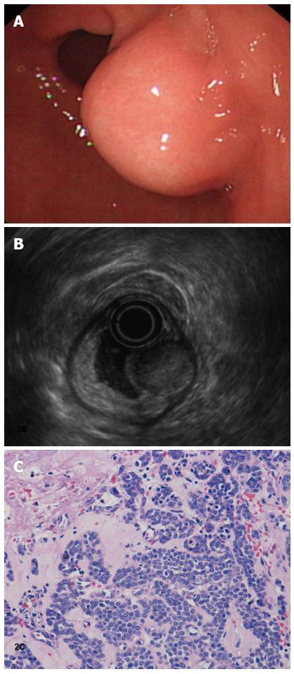
Figure 2 Gastroscopy shows a submucosa eminent lesion in the stomach wall.
A: Endoscopic ultrasonic shows a solid mass originating from the superficial layer of the muscular propria. It is 20 mm × 16 mm in size, and a marginal halo can be observed; B: The mass appears as a round, hypoechoic lesion with homogenous echogenicity. After a failed endoscopic submucosal dissection with ligation for the treatment of the small tumor, a subtotal gastrectomy was performed; C: Microscopic examination shows numerous dilated, thin-walled vascular spaces surrounded by uniform glomus cells (hematoxylin and eosin stain, × 50).
- Citation: Tang M, Hou J, Wu D, Han XY, Zeng MS, Yao XZ. Glomus tumor in the stomach: Computed tomography and endoscopic ultrasound findings. World J Gastroenterol 2013; 19(8): 1327-1329
- URL: https://www.wjgnet.com/1007-9327/full/v19/i8/1327.htm
- DOI: https://dx.doi.org/10.3748/wjg.v19.i8.1327


