Copyright
©2013 Baishideng Publishing Group Co.
World J Gastroenterol. Feb 28, 2013; 19(8): 1327-1329
Published online Feb 28, 2013. doi: 10.3748/wjg.v19.i8.1327
Published online Feb 28, 2013. doi: 10.3748/wjg.v19.i8.1327
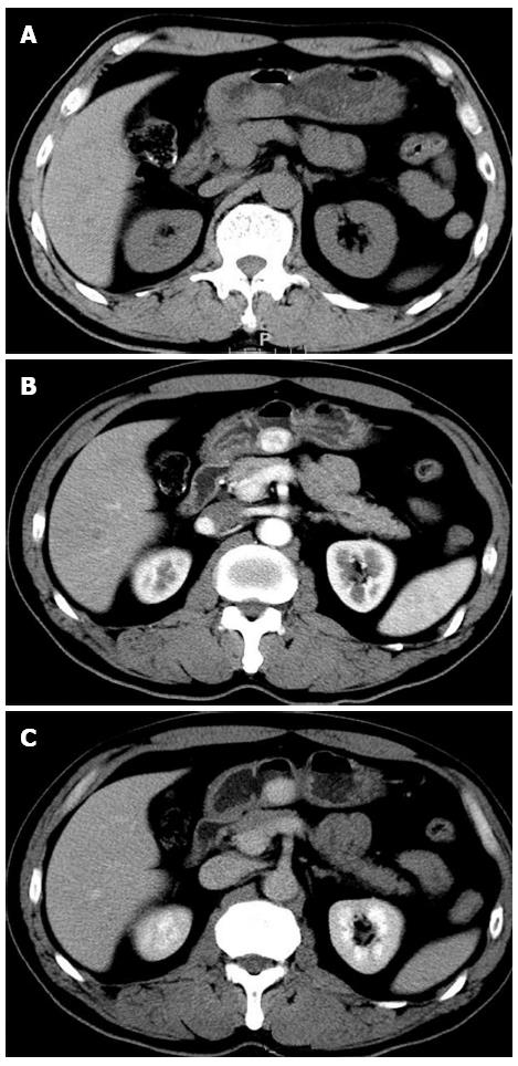
Figure 1 A 57-year-old man presented with intermittent dull abdominal pain after a duration of 1 year with a glomus tumor in the lesser curvature.
A: A plain computed tomography (CT) scan of the tumor revealing a clear regular contour and an isodensity mass; B: The mass shows dense homogeneous enhancement with an intact overlying mucosa on early-phase contrast-enhanced CT; C: The mass shows continuous homogeneous enhancement on delay-phase contrast-enhanced CT.
- Citation: Tang M, Hou J, Wu D, Han XY, Zeng MS, Yao XZ. Glomus tumor in the stomach: Computed tomography and endoscopic ultrasound findings. World J Gastroenterol 2013; 19(8): 1327-1329
- URL: https://www.wjgnet.com/1007-9327/full/v19/i8/1327.htm
- DOI: https://dx.doi.org/10.3748/wjg.v19.i8.1327


