Copyright
©2013 Baishideng Publishing Group Co.
World J Gastroenterol. Feb 28, 2013; 19(8): 1314-1317
Published online Feb 28, 2013. doi: 10.3748/wjg.v19.i8.1314
Published online Feb 28, 2013. doi: 10.3748/wjg.v19.i8.1314
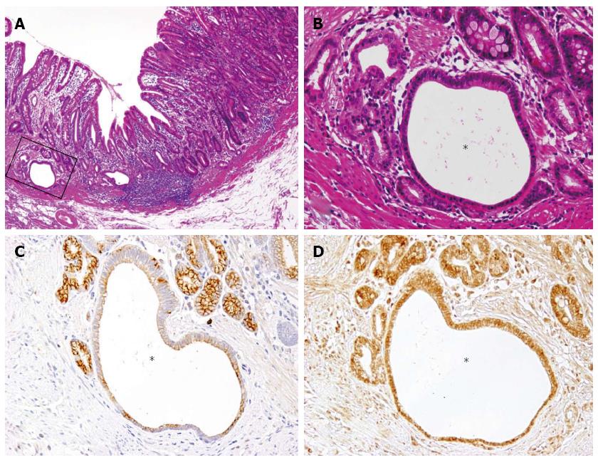
Figure 2 Expression of KCNE2 and estrogen receptor in non-neoplastic cystic lesion by immunohistochemistry.
A: Low power magnification of a transitional area from the non-neoplastic mucosal layer with intramucosal cystic lesions (left) to the intramucosal adenocarcinoma (right, HE, × 40); B: High power magnification (A, squared area) around the intramucosal cystic lesion (asterisk, HE, × 200); C: KCNE2 immunostaining of a serial section of (B) shows that KCNE2 is almost negative in the dilated cystic gland (asterisk), while the surrounding non-cystic glands are positive (× 200); D: Estrogen receptor immunostaining of serial sections of (B) and (C) show that ER is equally expressed in both cystic (asterisk) and non-cystic glands (× 200).
- Citation: Kuwahara N, Kitazawa R, Fujiishi K, Nagai Y, Haraguchi R, Kitazawa S. Gastric adenocarcinoma arising in gastritis cystica profunda presenting with selective loss of KCNE2 expression. World J Gastroenterol 2013; 19(8): 1314-1317
- URL: https://www.wjgnet.com/1007-9327/full/v19/i8/1314.htm
- DOI: https://dx.doi.org/10.3748/wjg.v19.i8.1314


