Copyright
©2013 Baishideng Publishing Group Co.
World J Gastroenterol. Feb 28, 2013; 19(8): 1152-1157
Published online Feb 28, 2013. doi: 10.3748/wjg.v19.i8.1152
Published online Feb 28, 2013. doi: 10.3748/wjg.v19.i8.1152
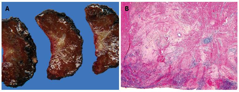
Figure 4 Pathological examination of the resected liver specimen corresponding to the lesion indicated by the white arrow in figure 3.
A: Gross specimen: on cut the macroscopic examination shows a star-like whitish lesion; B: At histology (hematoxylin and eosin, x 100) only fibrosis and inflammation are seen indicating a complete response to chemotherapy.
- Citation: Puppa G, Poston G, Jess P, Nash GF, Coenegrachts K, Stang A. Staging colorectal cancer with the TNM 7th: The presumption of innocence when applying the M category. World J Gastroenterol 2013; 19(8): 1152-1157
- URL: https://www.wjgnet.com/1007-9327/full/v19/i8/1152.htm
- DOI: https://dx.doi.org/10.3748/wjg.v19.i8.1152


