Copyright
©2013 Baishideng Publishing Group Co.
World J Gastroenterol. Feb 28, 2013; 19(8): 1152-1157
Published online Feb 28, 2013. doi: 10.3748/wjg.v19.i8.1152
Published online Feb 28, 2013. doi: 10.3748/wjg.v19.i8.1152
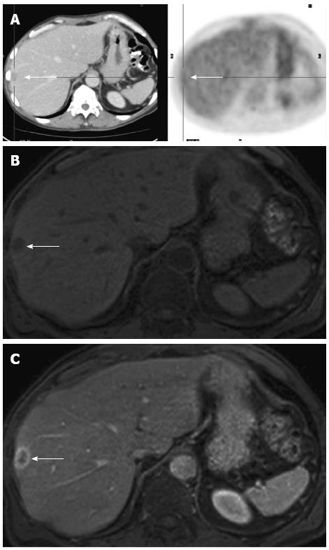
Figure 2 Example of a small lesion which proved to be malignant after follow-up.
A: Positron emission tomography (PET)/computed tomography negative (PET-cold) focal liver lesion (white arrow) in a patient who received chemotherapy in the past; B: Corresponding T1w Gradient Echo magnetic resonance imaging (MRI) image before injection of contrast agent shows a rather hypo-intense focal liver lesion (white arrow); C: Corresponding T1w Gradient Echo MRI image in the venous phase after injection of contrast agent shows a hypo-intense focal liver lesion with ring-enhancement compatible with an (active) malignant focal liver lesion (colorectal cancer liver metastasis) (white arrow).
- Citation: Puppa G, Poston G, Jess P, Nash GF, Coenegrachts K, Stang A. Staging colorectal cancer with the TNM 7th: The presumption of innocence when applying the M category. World J Gastroenterol 2013; 19(8): 1152-1157
- URL: https://www.wjgnet.com/1007-9327/full/v19/i8/1152.htm
- DOI: https://dx.doi.org/10.3748/wjg.v19.i8.1152


