Copyright
©2013 Baishideng Publishing Group Co.
World J Gastroenterol. Feb 28, 2013; 19(8): 1152-1157
Published online Feb 28, 2013. doi: 10.3748/wjg.v19.i8.1152
Published online Feb 28, 2013. doi: 10.3748/wjg.v19.i8.1152
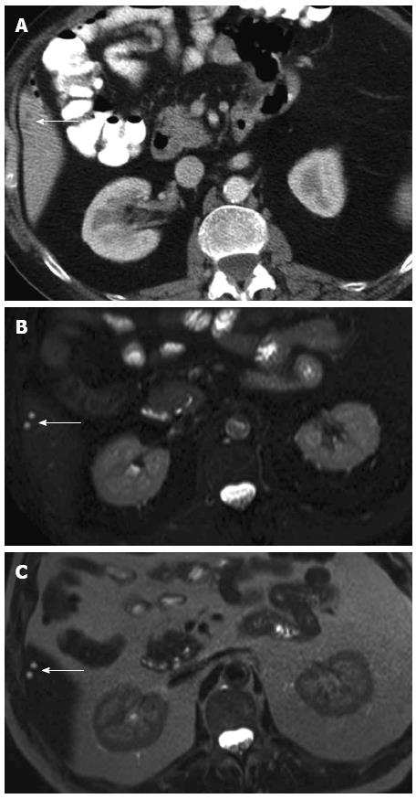
Figure 1 Example of a small lesion which proved to be benign after follow-up.
A: Two small (< 10 mm) indeterminate focal liver lesions (white arrow) on contrast-enhanced computed tomography; B: Corresponding magnetic resonance imaging (MRI), images showing a T2w Turbo Spin Echo sequence with fat suppression. Two clearly hyperintense focal liver lesions (white arrow) are displayed compatible with simple liver cysts; C: Corresponding MRI images showing a T2w Turbo Spin Echo sequence without fat suppression. Two clearly hyperintense focal liver lesions (white arrow) are displayed compatible with simple liver cysts.
- Citation: Puppa G, Poston G, Jess P, Nash GF, Coenegrachts K, Stang A. Staging colorectal cancer with the TNM 7th: The presumption of innocence when applying the M category. World J Gastroenterol 2013; 19(8): 1152-1157
- URL: https://www.wjgnet.com/1007-9327/full/v19/i8/1152.htm
- DOI: https://dx.doi.org/10.3748/wjg.v19.i8.1152


