Copyright
©2013 Baishideng Publishing Group Co.
World J Gastroenterol. Feb 21, 2013; 19(7): 1049-1055
Published online Feb 21, 2013. doi: 10.3748/wjg.v19.i7.1049
Published online Feb 21, 2013. doi: 10.3748/wjg.v19.i7.1049
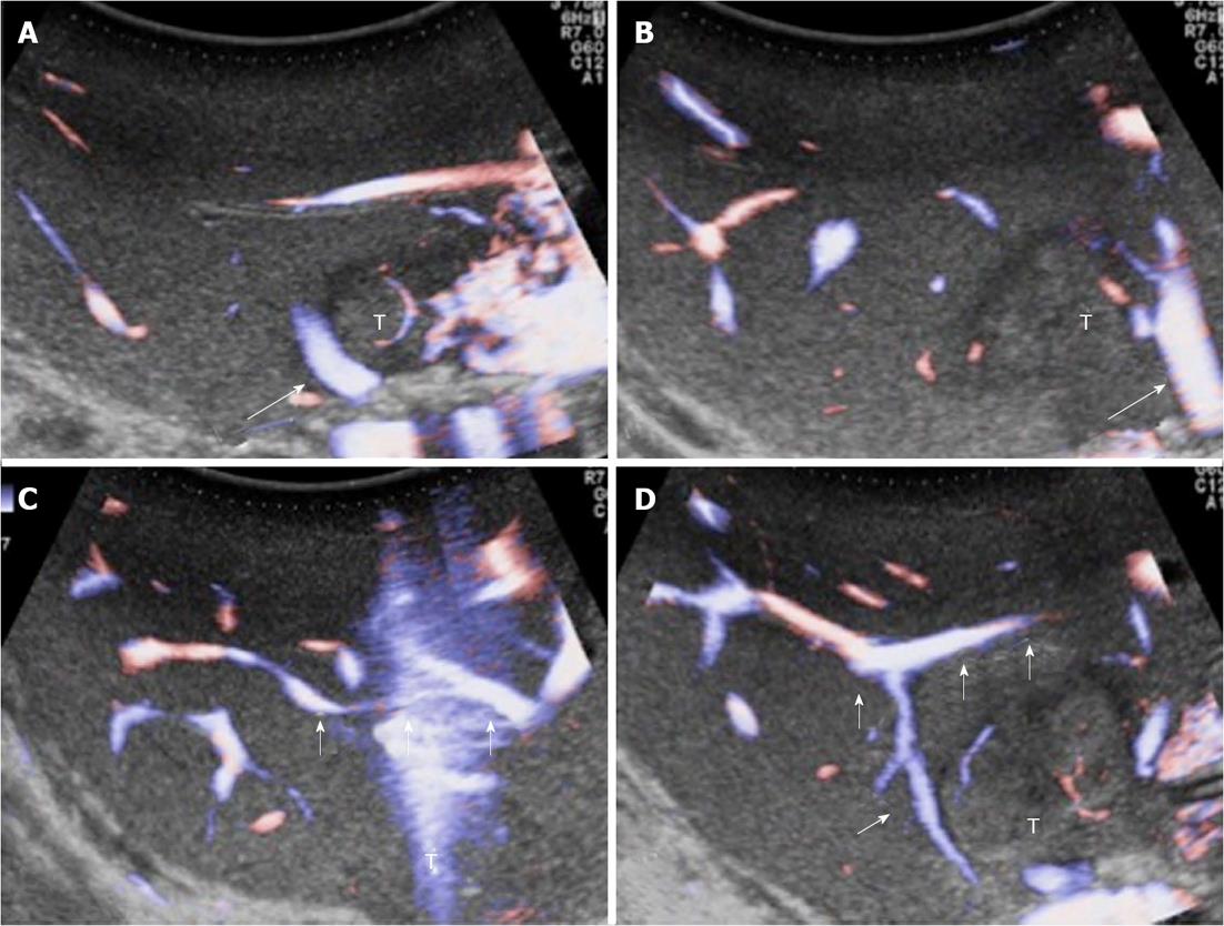
Figure 6 Intraoperative ultrasound study of communicating veins.
A: A tumour located between the middle hepatic vein (MHV) (arrow) and the left hepatic vein (LHV) at their confluence into the inferior vena cava; B: The arrow indicates the LHV; C, D: Evidence of communicating veins (arrows) between the LHV and the MHV. T: Tumor.
- Citation: Donadon M, Procopio F, Torzilli G. Tailoring the area of hepatic resection using inflow and outflow modulation. World J Gastroenterol 2013; 19(7): 1049-1055
- URL: https://www.wjgnet.com/1007-9327/full/v19/i7/1049.htm
- DOI: https://dx.doi.org/10.3748/wjg.v19.i7.1049


