Copyright
©2013 Baishideng Publishing Group Co.
World J Gastroenterol. Feb 14, 2013; 19(6): 951-954
Published online Feb 14, 2013. doi: 10.3748/wjg.v19.i6.951
Published online Feb 14, 2013. doi: 10.3748/wjg.v19.i6.951
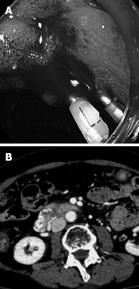
Figure 1 Endoscopy and computed tomography of the duodenum.
A: Endoscopy demonstrates bleeding varices in the second portion of the duodenum; B: Contrast-enhanced computed tomography reveals markedly tortuous varices around the wall in the second and third portion of the duodenum.
- Citation: Hashimoto R, Sofue K, Takeuchi Y, Shibamoto K, Arai Y. Successful balloon-occluded retrograde transvenous obliteration for bleeding duodenal varices using cyanoacrylate. World J Gastroenterol 2013; 19(6): 951-954
- URL: https://www.wjgnet.com/1007-9327/full/v19/i6/951.htm
- DOI: https://dx.doi.org/10.3748/wjg.v19.i6.951


