Copyright
©2013 Baishideng Publishing Group Co.
World J Gastroenterol. Feb 14, 2013; 19(6): 946-950
Published online Feb 14, 2013. doi: 10.3748/wjg.v19.i6.946
Published online Feb 14, 2013. doi: 10.3748/wjg.v19.i6.946
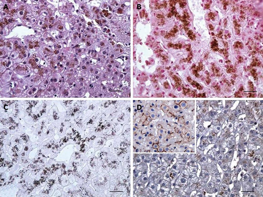
Figure 1 Histochemistry and immunohistology of the liver in Dubin-Johnson syndrome patient.
A: Accumulation of distinctive dark brown pigment in hepatocytes was detected in hematoxylin and eosin staining; B: The pigment was negative in Perls reaction; C: The pigment reduced Masson’s solution; D: Immunohistochemical analysis for ABCC2/MRP2 protein was negative compared to the positive control (inset). Original magnification ×400 (bars correspond to 100 μm).
- Citation: Sticova E, Elleder M, Hulkova H, Luksan O, Sauer M, Wunschova-Moudra I, Novotny J, Jirsa M. Dubin-Johnson syndrome coinciding with colon cancer and atherosclerosis. World J Gastroenterol 2013; 19(6): 946-950
- URL: https://www.wjgnet.com/1007-9327/full/v19/i6/946.htm
- DOI: https://dx.doi.org/10.3748/wjg.v19.i6.946


