Copyright
©2013 Baishideng Publishing Group Co.
World J Gastroenterol. Feb 14, 2013; 19(6): 855-865
Published online Feb 14, 2013. doi: 10.3748/wjg.v19.i6.855
Published online Feb 14, 2013. doi: 10.3748/wjg.v19.i6.855
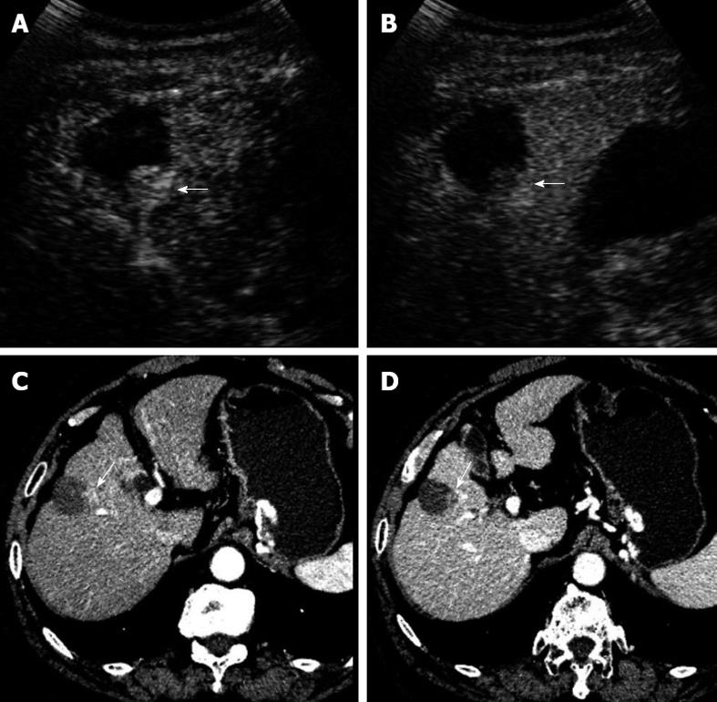
Figure 2 A 70-year-old male patient with hepatocellular carcinoma.
Local tumor progression (arrow) was detected 6 mos after radiofrequency ablation in combination with ethanol ablation for hepatocellular carcinoma in segment 5 of the liver. Local tumor progression showed hyper-enhancement in the arterial phase and iso-enhancement in the portal-late phase on contrast-enhanced ultrasound (A, B), whereas hyper-enhancement in the arterial phase and wash-out in the portal-venous phase on contrast-enhanced computed tomography (C, D).
- Citation: Zheng SG, Xu HX, Lu MD, Xie XY, Xu ZF, Liu GJ, Liu LN. Role of contrast-enhanced ultrasound in follow-up assessment after ablation for hepatocellular carcinoma. World J Gastroenterol 2013; 19(6): 855-865
- URL: https://www.wjgnet.com/1007-9327/full/v19/i6/855.htm
- DOI: https://dx.doi.org/10.3748/wjg.v19.i6.855


