Copyright
©2013 Baishideng Publishing Group Co.
World J Gastroenterol. Feb 7, 2013; 19(5): 692-705
Published online Feb 7, 2013. doi: 10.3748/wjg.v19.i5.692
Published online Feb 7, 2013. doi: 10.3748/wjg.v19.i5.692
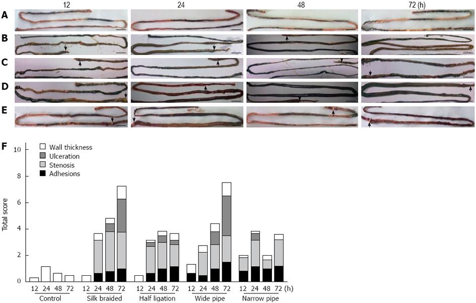
Figure 1 Dissection of the entire intestine from the pyloric antrum to the ileocecal valve in the obstructed and control rats.
A: Control; B: Braided silk; C: Half ligation; D: Wide pipe; E: Narrow pipe. Rats were killed at different time points after surgery; F: The morphological score of changes in the extent and severity of adhesions, stenosis, ulceration and bowel wall thickness. The arrow indicates the portion of obstructed intestine bearing a ring (scale bar: 1 cm).
- Citation: Yuan ML, Yang Z, Li YC, Shi LL, Guo JL, Huang YQ, Kang X, Cheng JJ, Chen Y, Yu T, Cao DQ, Pang H, Zhang X. Comparison of different methods of intestinal obstruction in a rat model. World J Gastroenterol 2013; 19(5): 692-705
- URL: https://www.wjgnet.com/1007-9327/full/v19/i5/692.htm
- DOI: https://dx.doi.org/10.3748/wjg.v19.i5.692


