Copyright
©2013 Baishideng Publishing Group Co.
World J Gastroenterol. Feb 7, 2013; 19(5): 682-691
Published online Feb 7, 2013. doi: 10.3748/wjg.v19.i5.682
Published online Feb 7, 2013. doi: 10.3748/wjg.v19.i5.682
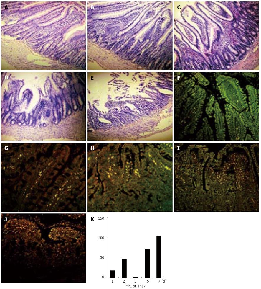
Figure 2 Expression levels of helper T cell 17 cells in intestine grafts paralleled with the degree of rejection in rat recipients.
The intestines from inbred F344 were transplanted to LEW rats. The recipient rats were sacrificed on the 1st, 2nd, 3rd, 5th, and 7th d after transplantation. The grafts were removed, fixed, and embedded. The sections were stained with hematoxylin eosin (HE), anti-rat-interleukin-17, and anti-rat-CD4 fluorescence antibodies (× 400). A-E: HE staining of intestine graft; F-J: Helper T cell 17 (Th17) immunofluorescence staining of intestine graft; A, F: 1 d after transplantation; B, G: 2 d after transplantation; C, H: 3 d after transplantation; D, I: 5 d after transplantation; E, J: 7 d after transplantation; K: Mean fluorescence intensity (MFI) of Th17 expression of intestine graft.
- Citation: Yang JJ, Feng F, Hong L, Sun L, Li MB, Zhuang R, Pan F, Wang YM, Wang WZ, Wu GS, Zhang HW. Interleukin-17 plays a critical role in the acute rejection of intestinal transplantation. World J Gastroenterol 2013; 19(5): 682-691
- URL: https://www.wjgnet.com/1007-9327/full/v19/i5/682.htm
- DOI: https://dx.doi.org/10.3748/wjg.v19.i5.682


