Copyright
©2013 Baishideng Publishing Group Co.
World J Gastroenterol. Feb 7, 2013; 19(5): 682-691
Published online Feb 7, 2013. doi: 10.3748/wjg.v19.i5.682
Published online Feb 7, 2013. doi: 10.3748/wjg.v19.i5.682
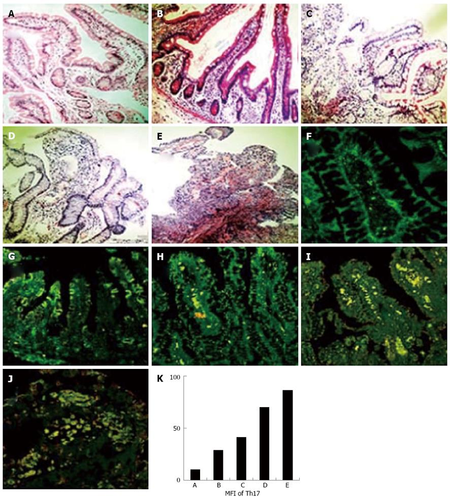
Figure 1 Expression levels of helper T cell 17 cells in intestine grafts paralleled with the degree of rejection in human recipients.
The paraffin-embedded human intestine tissues after small bowel transplantation were sectioned and stained with hematoxylin eosin (HE), anti-rat-interleukin-17, and anti-rat-CD4 fluorescence antibodies (× 400). A-E: HE staining of intestine graft; F-J: Helper T cell 17 (Th17) immunofluorescence staining of intestine graft; A, F: No acute rejection; B, G: Suspicious acute rejection; C, H: Mild acute rejection; D, I: Moderate acute rejection; E, J: Severe acute rejection; K: Mean fluorescence intensity (MFI) of Th17 expression of intestine graft.
- Citation: Yang JJ, Feng F, Hong L, Sun L, Li MB, Zhuang R, Pan F, Wang YM, Wang WZ, Wu GS, Zhang HW. Interleukin-17 plays a critical role in the acute rejection of intestinal transplantation. World J Gastroenterol 2013; 19(5): 682-691
- URL: https://www.wjgnet.com/1007-9327/full/v19/i5/682.htm
- DOI: https://dx.doi.org/10.3748/wjg.v19.i5.682


