Copyright
©2013 Baishideng Publishing Group Co.
World J Gastroenterol. Feb 7, 2013; 19(5): 673-681
Published online Feb 7, 2013. doi: 10.3748/wjg.v19.i5.673
Published online Feb 7, 2013. doi: 10.3748/wjg.v19.i5.673
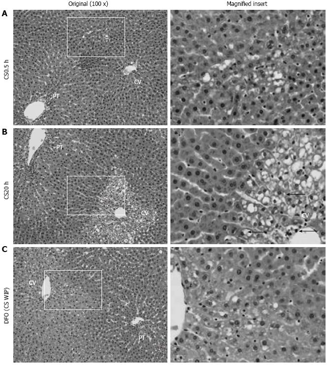
Figure 3 Histopathological appearance of livers following cold storage, warm ischemia and perfusion and the effect of desferrioxamine.
A: The control liver shows some early vacuolization in zone 2 following 0.5 h of cold storage (CS0.5) (magnified insert); B: Following 20 h of cold storage (CS20), the liver shows marked vacuolization and nuclear pyknosis in zones 3 as indicated by arrows (←, magnified insert); C: Following 20 h of CS with desferrioxamine (DFO) in CS, warm ischemia and perfusion media (CS WIP), the liver shows mild vacuolization in zone 3 (magnified insert). Hematoxylin and eosin staining and the original magnification was 100 ×. PT: Portal tract; CV: Central vein.
- Citation: Arthur PG, Niu XW, Huang WH, DeBoer B, Lai CT, Rossi E, Joseph J, Jeffrey GP. Desferrioxamine in warm reperfusion media decreases liver injury aggravated by cold storage. World J Gastroenterol 2013; 19(5): 673-681
- URL: https://www.wjgnet.com/1007-9327/full/v19/i5/673.htm
- DOI: https://dx.doi.org/10.3748/wjg.v19.i5.673


