Copyright
©2013 Baishideng Publishing Group Co.
World J Gastroenterol. Dec 28, 2013; 19(48): 9490-9494
Published online Dec 28, 2013. doi: 10.3748/wjg.v19.i48.9490
Published online Dec 28, 2013. doi: 10.3748/wjg.v19.i48.9490
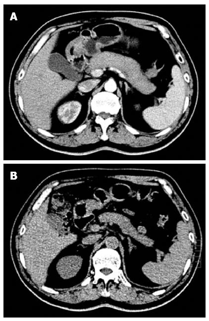
Figure 1 Typical imaging features of type 1 autoimmune pancreatitis.
Computed tomography (CT) scan showing diffuse swelling of the pancreas with loss of lobulation (A), and a dramatic decrease in swelling of the pancreas after 3 wk of steroid treatment (B).
- Citation: Qu LM, Liu YH, Brigstock DR, Wen XY, Liu YF, Li YJ, Gao RP. IgG4-related autoimmune pancreatitis overlapping with Mikulicz’s disease and lymphadenitis: A case report. World J Gastroenterol 2013; 19(48): 9490-9494
- URL: https://www.wjgnet.com/1007-9327/full/v19/i48/9490.htm
- DOI: https://dx.doi.org/10.3748/wjg.v19.i48.9490


