Copyright
©2013 Baishideng Publishing Group Co.
World J Gastroenterol. Dec 28, 2013; 19(48): 9485-9489
Published online Dec 28, 2013. doi: 10.3748/wjg.v19.i48.9485
Published online Dec 28, 2013. doi: 10.3748/wjg.v19.i48.9485
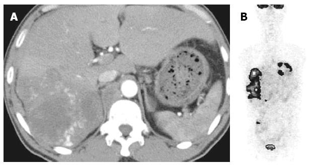
Figure 1 Enhanced computed tomography of the liver and fludeoxyglucose-positron emission tomography.
A: Enhanced computed tomography showed a multi-nodular tumor in the right lobe of the liver; B: Fludeoxyglucose-positron emission tomography scan showed accumulation in the liver with no accumulation in the testes.
- Citation: Sekine R, Hyodo M, Kojima M, Meguro Y, Suzuki A, Yokoyama T, Lefor AT, Hirota N. Primary hepatic choriocarcinoma in a 49-year-old man: Report of a case. World J Gastroenterol 2013; 19(48): 9485-9489
- URL: https://www.wjgnet.com/1007-9327/full/v19/i48/9485.htm
- DOI: https://dx.doi.org/10.3748/wjg.v19.i48.9485


