Copyright
©2013 Baishideng Publishing Group Co.
World J Gastroenterol. Dec 28, 2013; 19(48): 9481-9484
Published online Dec 28, 2013. doi: 10.3748/wjg.v19.i48.9481
Published online Dec 28, 2013. doi: 10.3748/wjg.v19.i48.9481
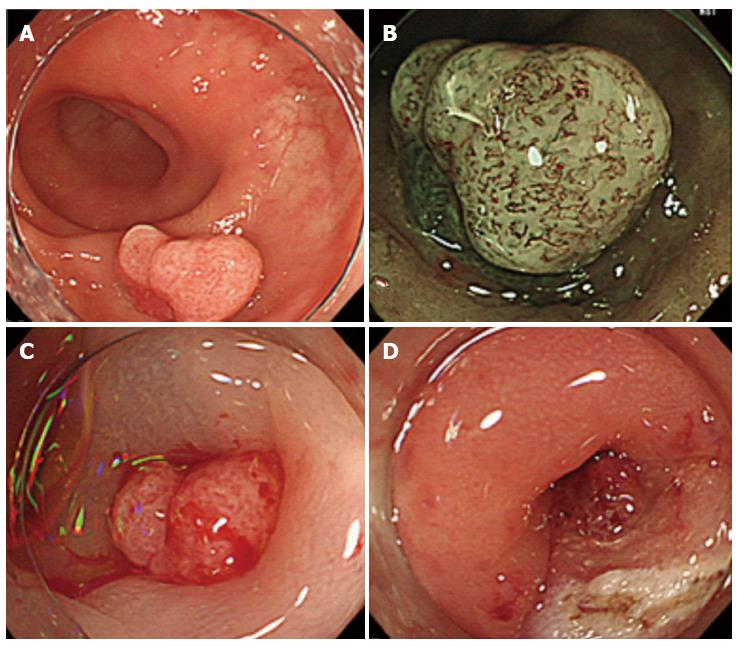
Figure 1 Endoscopic mucosal dissection of the sigmoid colon polyp.
A: A sigmoid colon polyp approximately 25 mm in diameter; B: Narrow band imaging magnified colonoscopy was performed to investigate the polyp in greater detail. Several irregular microvessels were observed on the surface of the polyp, but there was no pit pattern on the surface; C: A local saline injection was administered, and we observed slight elevation of the polyp; D: After the endoscopic mucosal resection procedure and the removal of the polyp, the diverticulum was identified using the resected stalk of the polyp.
- Citation: Mori H, Tsushimi T, Kobara H, Nishiyama N, Fujihara S, Matsunaga T, Ayagi M, Yachida T, Masaki T. Endoscopic management of a rare granulation polyp in a colonic diverticulum. World J Gastroenterol 2013; 19(48): 9481-9484
- URL: https://www.wjgnet.com/1007-9327/full/v19/i48/9481.htm
- DOI: https://dx.doi.org/10.3748/wjg.v19.i48.9481


