Copyright
©2013 Baishideng Publishing Group Co.
World J Gastroenterol. Dec 28, 2013; 19(48): 9334-9342
Published online Dec 28, 2013. doi: 10.3748/wjg.v19.i48.9334
Published online Dec 28, 2013. doi: 10.3748/wjg.v19.i48.9334
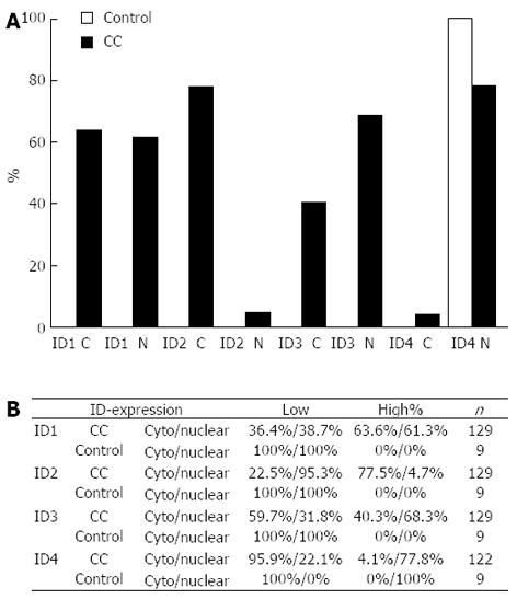
Figure 3 Proportion of inhibitor of differentiation protein expression in biliary tract cancer and normal intrahepatic bile ducts.
A: Percentage of biliary tract cancer (BTC) with high expression of the respective inhibitor of differentiation (ID) protein in BTC (black bars) and normal intrahepatic bile ducts (control; grey bar); B: Summary of ID protein expression. Overall ID protein expression ID protein expression was scored in low (0+1) and high (2+3) and assessed for nuclear and cytoplasmic (cyto) staining pattern. Cytoplasmic staining intensity was scored 0 (negative) to 3 (strong), nuclear expression was scored based on the percentage of positive nuclei (0: 0%-10%; 1: 11%-50%; 2: 51%-80%; and 3: 81%-100%). For ID4, only 122 samples could be analyzed. C: Cytoplasmic expression; N: Nuclear expression.
- Citation: Harder J, Müller MJ, Fuchs M, Gumpp V, Schmitt-Graeff A, Fischer R, Frank M, Opitz O, Hasskarl J. Inhibitor of differentiation proteins do not influence prognosis of biliary tract cancer. World J Gastroenterol 2013; 19(48): 9334-9342
- URL: https://www.wjgnet.com/1007-9327/full/v19/i48/9334.htm
- DOI: https://dx.doi.org/10.3748/wjg.v19.i48.9334


