Copyright
©2013 Baishideng Publishing Group Co.
World J Gastroenterol. Dec 21, 2013; 19(47): 9127-9132
Published online Dec 21, 2013. doi: 10.3748/wjg.v19.i47.9127
Published online Dec 21, 2013. doi: 10.3748/wjg.v19.i47.9127
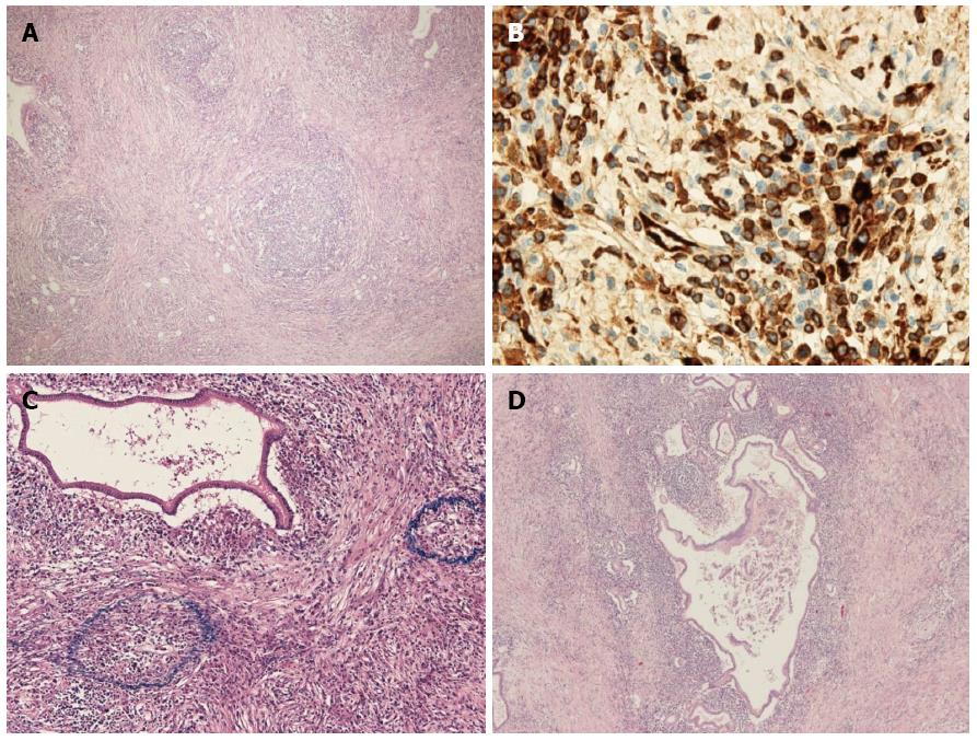
Figure 6 Microscopic findings.
A: Each solid lesion presented a striform pattern with lymphoid follicles and inflammatory cells (HE, original magnification × 12.5); B: The plasma cells showed positivity for immunoglobulin G4 (HE, original magnification × 200); C: Many obliterating phlebitides were observed (HE, original magnification × 100); D: The multilocular cysts produced mucus and demonstrated a papillary pattern (HE, original magnification × 12.5).
- Citation: Urata T, Naito Y, Izumi Y, Takekuma Y, Yokomizo H, Nagamine M, Fukuda S, Notohara K, Hifumi M. Localized type 1 autoimmune pancreatitis superimposed upon preexisting intraductal papillary mucinous neoplasms. World J Gastroenterol 2013; 19(47): 9127-9132
- URL: https://www.wjgnet.com/1007-9327/full/v19/i47/9127.htm
- DOI: https://dx.doi.org/10.3748/wjg.v19.i47.9127


