Copyright
©2013 Baishideng Publishing Group Co.
World J Gastroenterol. Dec 21, 2013; 19(47): 9092-9103
Published online Dec 21, 2013. doi: 10.3748/wjg.v19.i47.9092
Published online Dec 21, 2013. doi: 10.3748/wjg.v19.i47.9092
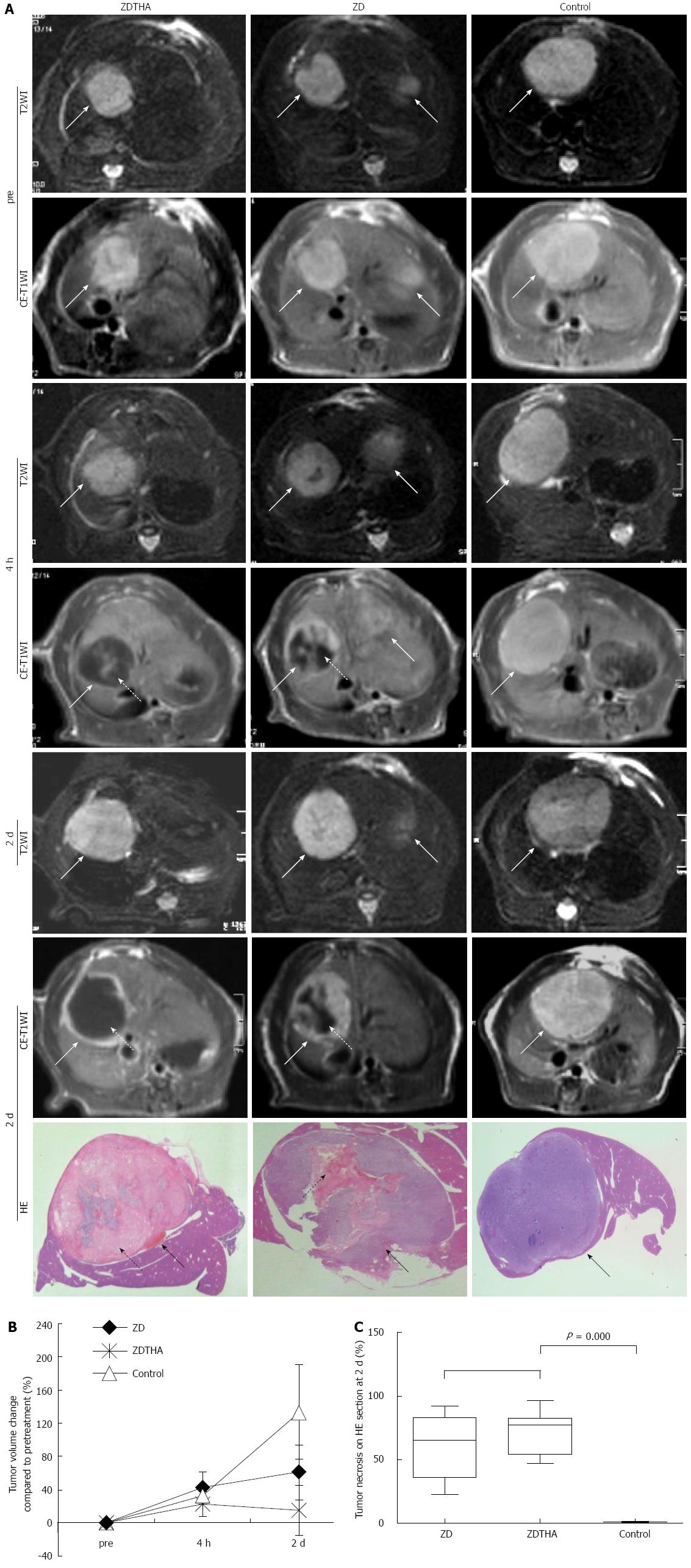
Figure 1 Tumor growth delay.
A: Representative axial magnetic resonance images of liver tumors acquired with T2-weighted images (T2WI) [repetition/echo time (TR/TE) = 3860/106 ms], contrast enhanced T1-weighted images (CE-T1WI) (TR/TE = 535/9.2 ms) and HE stained sections. Top row magnetic resonance images: After the combination therapy with ZDTHA, the tumor (solid arrow) in the right liver lobe showed delayed growth with massive central necrotic area (dotted arrow) compared to the control tumor on day 2; Middle row magnetic resonance images: After ZD6126 treatment, the right tumor also showed delayed growth compared to the control tumor on day 2 , however, the tumor necrotic area was reduced (dotted arrow) because the tumor regrew from viable rim; Bottom row magnetic resonance images: In a control animal, the tumor grew remarkably at 2 d; HE sections: The tumor (solid arrow) and central necrotic areas (dotted arrow) in different groups were verified by HE staining; B: The graph indicated that ZDTHA induced a significant tumor volume growth delay during the experiment, compared to both the ZD6126 and control groups (bP < 0.01 for both; C: The box plots showed significantly higher percentages of necrotic area (necrosis/tumor) on HE stained sections in both the ZDTHA and ZD6126 groups compared to the control group (P = 0.000 for both). No significant difference in necrosis was found between the ZDTHA and ZD6126 groups (P = 0.09).
- Citation: Chen F, Keyzer FD, Feng YB, Cona MM, Yu J, Marchal G, Oyen R, Ni YC. Separate calculation of DW-MRI in assessing therapeutic effect in liver tumors in rats. World J Gastroenterol 2013; 19(47): 9092-9103
- URL: https://www.wjgnet.com/1007-9327/full/v19/i47/9092.htm
- DOI: https://dx.doi.org/10.3748/wjg.v19.i47.9092


