Copyright
©2013 Baishideng Publishing Group Co.
World J Gastroenterol. Dec 21, 2013; 19(47): 9034-9042
Published online Dec 21, 2013. doi: 10.3748/wjg.v19.i47.9034
Published online Dec 21, 2013. doi: 10.3748/wjg.v19.i47.9034
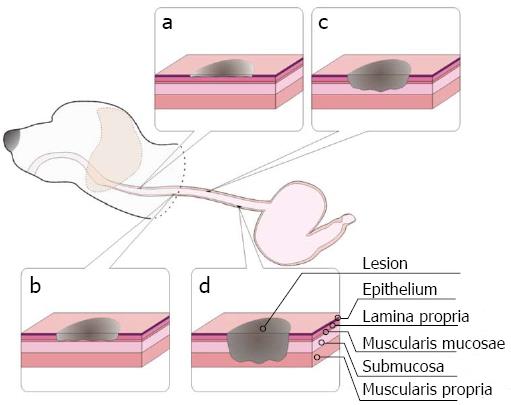
Figure 2 Schematic diagrams of superficial lesions in the esophagus with different depths in a canine model.
- Citation: Li JJ, He LJ, Shan HB, Wang TD, Xiong H, Chen LM, Xu GL, Li XH, Huang XX, Luo GY, Li Y, Zhang R. Superficial esophageal lesions detected by endoscopic ultrasound enhanced with submucosal edema. World J Gastroenterol 2013; 19(47): 9034-9042
- URL: https://www.wjgnet.com/1007-9327/full/v19/i47/9034.htm
- DOI: https://dx.doi.org/10.3748/wjg.v19.i47.9034


