Copyright
©2013 Baishideng Publishing Group Co.
World J Gastroenterol. Dec 21, 2013; 19(47): 9003-9011
Published online Dec 21, 2013. doi: 10.3748/wjg.v19.i47.9003
Published online Dec 21, 2013. doi: 10.3748/wjg.v19.i47.9003
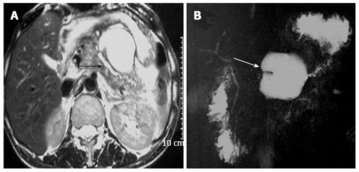
Figure 4 Magnetic resonance images.
T2 weighted axial image (A) and magnetic resonance cholangiopancreatography (B) in a case of traumatic pancreatitis show heterogenous signal intensity of pancreas with peripancreatic stranding. Main pancreatic duct is dilated in the body and tail region (black arrow). A lobulated pseudopancreatic cyst is seen in lesser sac anterior aspect of body of pancreas (white arrow) demonstrated in magnetic resonance cholangiopancreatography.
- Citation: Debi U, Kaur R, Prasad KK, Sinha SK, Sinha A, Singh K. Pancreatic trauma: A concise review. World J Gastroenterol 2013; 19(47): 9003-9011
- URL: https://www.wjgnet.com/1007-9327/full/v19/i47/9003.htm
- DOI: https://dx.doi.org/10.3748/wjg.v19.i47.9003


