Copyright
©2013 Baishideng Publishing Group Co.
World J Gastroenterol. Dec 14, 2013; 19(46): 8793-8798
Published online Dec 14, 2013. doi: 10.3748/wjg.v19.i46.8793
Published online Dec 14, 2013. doi: 10.3748/wjg.v19.i46.8793
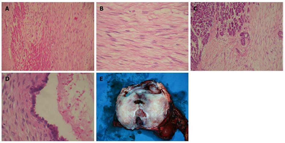
Figure 9 Pathological examination.
A: Pancreatic desmoid tumor with invasion to the duodenal wall; B: Proliferation of spindle-shaped stellate cells in fasciculate and storiform growth patterns within a myxoid intercellular matrix; C: Pancreatic infiltration by the tumor; D: The cystic area resulted from dilatation of entrapped excretory pancreatic ducts; E: Gross view of resected tumor and pancreas.
- Citation: Xu B, Zhu LH, Wu JG, Wang XF, Matro E, Ni JJ. Pancreatic solid cystic desmoid tumor: Case report and literature review. World J Gastroenterol 2013; 19(46): 8793-8798
- URL: https://www.wjgnet.com/1007-9327/full/v19/i46/8793.htm
- DOI: https://dx.doi.org/10.3748/wjg.v19.i46.8793


