Copyright
©2013 Baishideng Publishing Group Co.
World J Gastroenterol. Dec 14, 2013; 19(46): 8793-8798
Published online Dec 14, 2013. doi: 10.3748/wjg.v19.i46.8793
Published online Dec 14, 2013. doi: 10.3748/wjg.v19.i46.8793
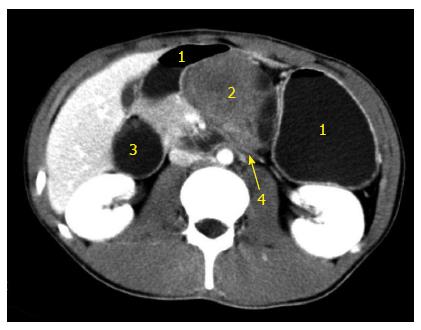
Figure 3 Abdominal computed tomography with contrast (venous phase) demonstrating a solid cystic mass invading the horizontal portion of the duodenum.
1: Stomach; 2: Pancreatic cystic mass; 3: Enlarged duodenum; 4: Horizontal duodenal portion invaded by the tumor.
- Citation: Xu B, Zhu LH, Wu JG, Wang XF, Matro E, Ni JJ. Pancreatic solid cystic desmoid tumor: Case report and literature review. World J Gastroenterol 2013; 19(46): 8793-8798
- URL: https://www.wjgnet.com/1007-9327/full/v19/i46/8793.htm
- DOI: https://dx.doi.org/10.3748/wjg.v19.i46.8793


