Copyright
©2013 Baishideng Publishing Group Co.
World J Gastroenterol. Dec 7, 2013; 19(45): 8449-8452
Published online Dec 7, 2013. doi: 10.3748/wjg.v19.i45.8449
Published online Dec 7, 2013. doi: 10.3748/wjg.v19.i45.8449
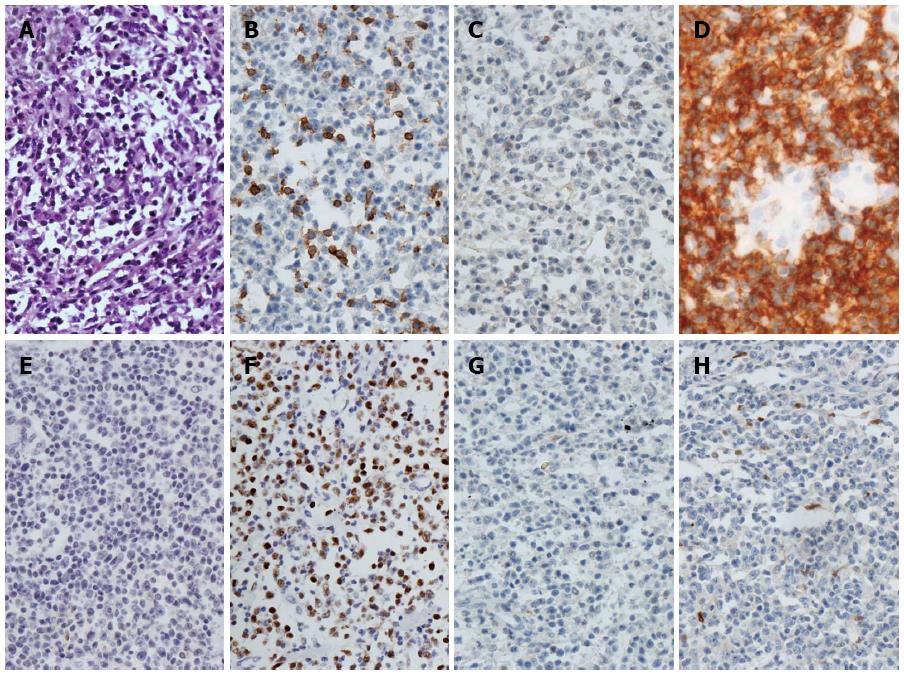
Figure 4 Histological and immunohistological examination of the endoscopic and surgical specimens showing diffuse large B-cell non-Hodgkin’s lymphoma.
A: × 400, HE staining; B: × 400, CD5 (-); C: × 400, CD10 (-); D: × 400, CD20 (+); E: × 400, CD23 (-); F: × 400, MUM-1 (+); G: × 400, Bcl-6 (-); H: × 400, Cyclin D1 (-).
- Citation: Xu XQ, Hong T, Li BL, Liu W. Ileo-ileal intussusception caused by diffuse large B-cell lymphoma of the ileum. World J Gastroenterol 2013; 19(45): 8449-8452
- URL: https://www.wjgnet.com/1007-9327/full/v19/i45/8449.htm
- DOI: https://dx.doi.org/10.3748/wjg.v19.i45.8449


