Copyright
©2013 Baishideng Publishing Group Co.
World J Gastroenterol. Dec 7, 2013; 19(45): 8440-8444
Published online Dec 7, 2013. doi: 10.3748/wjg.v19.i45.8440
Published online Dec 7, 2013. doi: 10.3748/wjg.v19.i45.8440
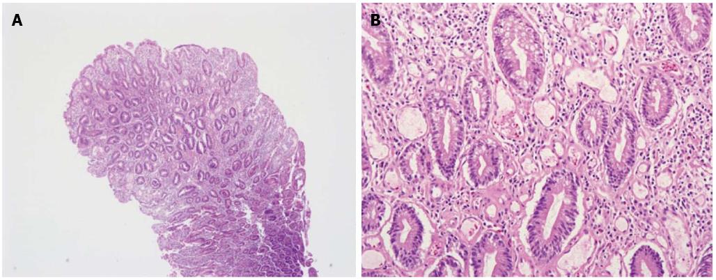
Figure 3 Histopathologic findings of intestinal lymphangiectasia.
A, B: Microscopic examination shows dilated lymphatic channels in the lamina propria (hematoxylin and eosin, × 40). Protein-rich fluid can escape from these channels into the extracellular space of the lamina propria and ultimately into the gut lumen (hematoxylin and eosin, × 200).
- Citation: Park MS, Lee BJ, Gu DH, Pyo JH, Kim KJ, Lee YH, Joo MK, Park JJ, Kim JS, Bak YT. Ileal polypoid lymphangiectasia bleeding diagnosed and treated by double balloon enteroscopy. World J Gastroenterol 2013; 19(45): 8440-8444
- URL: https://www.wjgnet.com/1007-9327/full/v19/i45/8440.htm
- DOI: https://dx.doi.org/10.3748/wjg.v19.i45.8440


