Copyright
©2013 Baishideng Publishing Group Co.
World J Gastroenterol. Dec 7, 2013; 19(45): 8435-8439
Published online Dec 7, 2013. doi: 10.3748/wjg.v19.i45.8435
Published online Dec 7, 2013. doi: 10.3748/wjg.v19.i45.8435
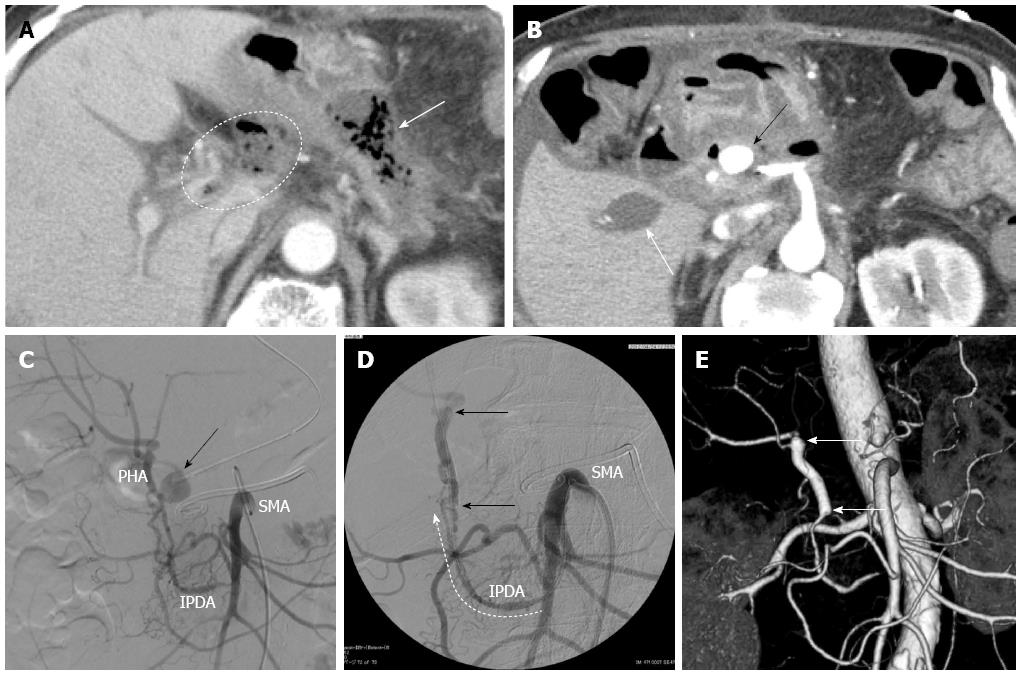
Figure 2 Imaging findings before and after endovascular treatment for the pseudoaneurysm.
A: Computed tomography (CT) image indicating formation of an intra-abdominal abscess around the remnant pancreas head (arrow) with an occluded portal vein due to the spreading inflammation around the abscess (dotted circle); B: CT image showing the pseudoaneurysm emerging from the common hepatic artery (CHA) stump (black arrow). The white arrow shows the occluded right portal vein; C: Angiography image confirming the pseudoaneurysm emerging from the CHA stump (arrow); D: Covered stent placement (arrows) performed via the IPDA (dotted arrow) originating from the superior mesenteric artery (SMA); E: CT angiography image at the 6-mo follow-up showing the patent covered stent (arrows) and sustained hepatic artery flow. IPDA: inferior pancreaticoduodenal artery; PHA: proper hepatic artery.
- Citation: Sumiyoshi T, Shima Y, Noda Y, Hosoki S, Hata Y, Okabayashi T, Kozuki A, Nakamura T. Endovascular pseudoaneurysm repair after distal pancreatectomy with celiac axis resection. World J Gastroenterol 2013; 19(45): 8435-8439
- URL: https://www.wjgnet.com/1007-9327/full/v19/i45/8435.htm
- DOI: https://dx.doi.org/10.3748/wjg.v19.i45.8435


