Copyright
©2013 Baishideng Publishing Group Co.
World J Gastroenterol. Dec 7, 2013; 19(45): 8269-8281
Published online Dec 7, 2013. doi: 10.3748/wjg.v19.i45.8269
Published online Dec 7, 2013. doi: 10.3748/wjg.v19.i45.8269
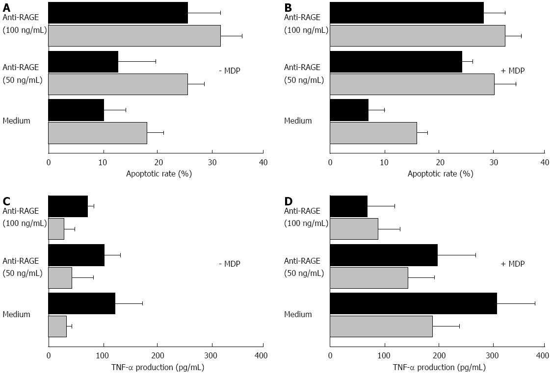
Figure 8 Functional assays.
The in vitro apoptotic rates of lamina propria mononuclear cells (LPMC) incubated in the absence or presence of two different concentrations of the anti-receptor for the advanced glycation end products (RAGE) blocking antibody, and with or without the muramyl dipeptide (MDP) used as antigenic stimulation are given in panels A and B. The analysis was carried out by flow cytometry, and the mean percentage values ± SD of at least three experiments for each condition were the following: in the absence of MDP (A), LPMC from non-diseased mucosa (grey bars) showed a spontaneous apoptotic rate of 18.4 ± 3.1, and a value of 26.9 ± 2.8 and 32.7 ± 4.6 in the presence of 50 and 100 ng/mL concentration of the anti-RAGE blocking antibody, respectively; when using LPMC from diseased mucosa (black bars), a value of spontaneous apoptotic rate of 10.1 ± 4.2 was found, while when incubating with 50 and 100 ng/mL concentration of the anti-RAGE blocking antibody, values of 13.0 ± 6.2 and 26.1 ± 5.5 were found, respectively. In the presence of MDP (B), LPMC from non-diseased mucosa showed a spontaneous apoptotic rate of 15.6 ± 2.1, and values of 31.9 ± 3.8 and 33.3 ± 2.9 in the presence of 50 and 100 ng/mL concentration of the anti-RAGE blocking antibody, respectively; when using LPMC from diseased mucosa, a value of spontaneous apoptotic rate of 7.8 ± 2.6 was found, while when incubating with 50 and 100 ng/mL concentration of the anti-RAGE blocking antibody, values of 24.1 ± 2.2 and 28.0 ± 4.1, respectively, were found. The tumor necrosis factor (TNF)-α production of LPMC cultured in vitro in the absence or presence of two different concentrations of the anti-RAGE blocking antibody, and with or without the MDP as antigenic stimulation are given in the panels C and D. The analysis was carried out by ELISA assay on culture supernatants, and the mean values ± SD were as follows: in the absence of MDP (C), the cytokine level was 32 ± 14 pg/mL in the cultures with LPMC from non-diseased areas, and 124 ± 48 pg/mL in those with LPMC from diseased areas; when incubating with 50 and 100 ng/mL of the anti-RAGE blocking antibody, values of 41 ± 38 and 27 ± 21 pg/mL for LPMC from non-diseased areas, and of 102 ± 29 and 67 ± 11 pg/mL for LPMC from diseased areas, respectively, were observed. In the presence of MDP (D), the TNF-α level was 184 ± 49 pg/mL in the cultures with LPMC from non-diseased areas, and 307 ± 68 pg/mL in those with LPMC from diseased areas; when incubating with 50 and 100 ng/mL of the anti-RAGE blocking antibody, values of 138 ± 50 and 87 ± 41 pg/mL for LPMC from non-diseased areas, and of 198 ± 68 and 71 ± 47 pg/mL for LPMC from diseased areas, respectively, were observed.
- Citation: Ciccocioppo R, Vanoli A, Klersy C, Imbesi V, Boccaccio V, Manca R, Betti E, Cangemi GC, Strada E, Besio R, Rossi A, Falcone C, Ardizzone S, Fociani P, Danelli P, Corazza GR. Role of the advanced glycation end products receptor in Crohn’s disease inflammation. World J Gastroenterol 2013; 19(45): 8269-8281
- URL: https://www.wjgnet.com/1007-9327/full/v19/i45/8269.htm
- DOI: https://dx.doi.org/10.3748/wjg.v19.i45.8269


