Copyright
©2013 Baishideng Publishing Group Co.
World J Gastroenterol. Nov 28, 2013; 19(44): 8156-8159
Published online Nov 28, 2013. doi: 10.3748/wjg.v19.i44.8156
Published online Nov 28, 2013. doi: 10.3748/wjg.v19.i44.8156
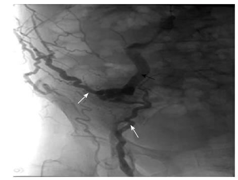
Figure 3 Opacification showed the varices fed by the superior mesenteric vein (black arrow) and communicated to the paraumbilical vein and femoral vein (white arrows).
- Citation: Yao DH, Luo XF, Zhou B, Li X. Ileal conduit stomal variceal bleeding managed by endovascular embolization. World J Gastroenterol 2013; 19(44): 8156-8159
- URL: https://www.wjgnet.com/1007-9327/full/v19/i44/8156.htm
- DOI: https://dx.doi.org/10.3748/wjg.v19.i44.8156


