Copyright
©2013 Baishideng Publishing Group Co.
World J Gastroenterol. Nov 21, 2013; 19(43): 7816-7819
Published online Nov 21, 2013. doi: 10.3748/wjg.v19.i43.7816
Published online Nov 21, 2013. doi: 10.3748/wjg.v19.i43.7816
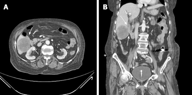
Figure 1 Initial abdominopelvic computed tomography findings.
Enhanced computed tomography shows a 5.7 cm × 5.2 cm heterogeneous enhancing liver mass connected with a small mass of the distal ileum (arrows). A: Axial view; B: Coronal view.
- Citation: Lee YH, Koo JS, Jung CH, Chung SY, Lee JJ, Kim SY, Hyun JJ, Jung SW, Choung RS, Lee SW, Choi JH. Development of enterohepatic fistula after embolization in ileal gastrointestinal stromal tumor: A case report. World J Gastroenterol 2013; 19(43): 7816-7819
- URL: https://www.wjgnet.com/1007-9327/full/v19/i43/7816.htm
- DOI: https://dx.doi.org/10.3748/wjg.v19.i43.7816


