Copyright
©2013 Baishideng Publishing Group Co.
World J Gastroenterol. Nov 21, 2013; 19(43): 7680-7695
Published online Nov 21, 2013. doi: 10.3748/wjg.v19.i43.7680
Published online Nov 21, 2013. doi: 10.3748/wjg.v19.i43.7680
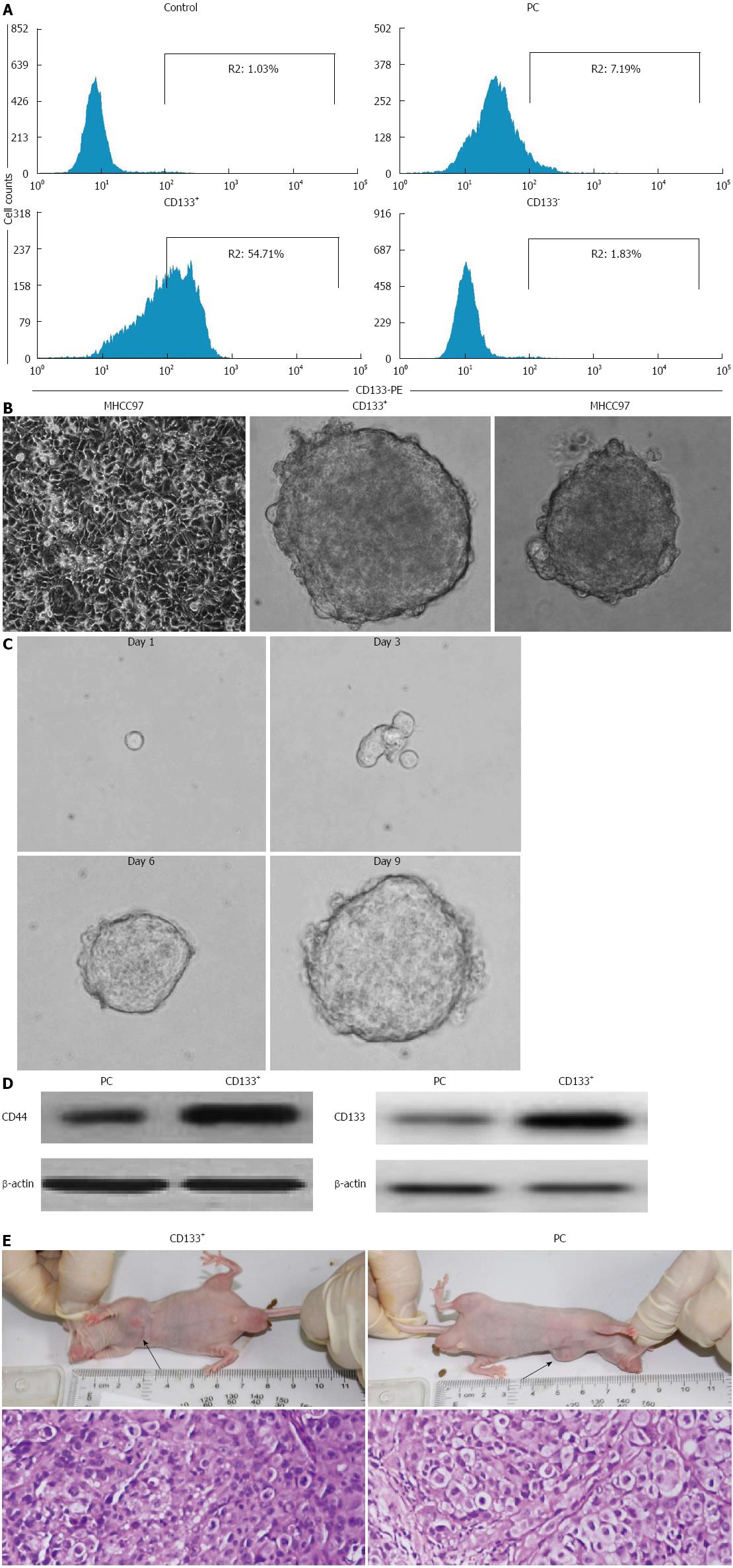
Figure 1 Isolation and characterization of liver cancer stem cells derived from the MHCC97 cell line.
A: Flow cytometry analysis of CD133 expression following sorting. CD133+ cells from MHCC97 cells formed liver cancer spheroids in stem cell-conditioned medium (200 × magnification); B: Anchorage-dependent growth of MHCC97 cells, tumor spheroid formed by CD133+ cells, tumor spheroid formed by parental MHCC97 cells; C: Secondary tumorspheres formed by single cells from dissociated primary liver spheroids (400 × magnification); D: Expression of stem cell surface markers CD44 and CD133 in CD133+ sphere-forming cells (SFCs) and parental cells; E: Hematoxylin-eosin staining revealed similar histological characteristics in tumor xenografts derived from CD133+ SFCs and their parental cells (100 × magnification).
-
Citation: Quan MF, Xiao LH, Liu ZH, Guo H, Ren KQ, Liu F, Cao JG, Deng XY. 8-bromo-7-methoxychrysin inhibits properties of liver cancer stem cells
via downregulation of β-catenin. World J Gastroenterol 2013; 19(43): 7680-7695 - URL: https://www.wjgnet.com/1007-9327/full/v19/i43/7680.htm
- DOI: https://dx.doi.org/10.3748/wjg.v19.i43.7680


