Copyright
©2013 Baishideng Publishing Group Co.
World J Gastroenterol. Nov 21, 2013; 19(43): 7661-7670
Published online Nov 21, 2013. doi: 10.3748/wjg.v19.i43.7661
Published online Nov 21, 2013. doi: 10.3748/wjg.v19.i43.7661
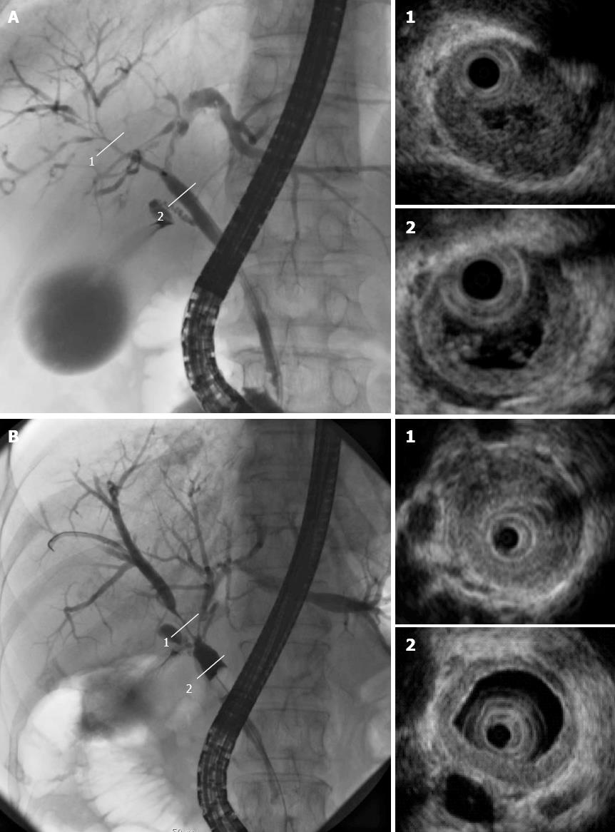
Figure 3 Cholangiogram displaying stenosis in the intrahepatic ducts (A-1) and hilar hepatic lesions (B-1); intraductal ultrasonography revealing bile duct wall thickening in areas with stenosis (1) and without (2).
- Citation: Nakazawa T, Naitoh I, Hayashi K, Miyabe K, Simizu S, Joh T. Diagnosis of IgG4-related sclerosing cholangitis. World J Gastroenterol 2013; 19(43): 7661-7670
- URL: https://www.wjgnet.com/1007-9327/full/v19/i43/7661.htm
- DOI: https://dx.doi.org/10.3748/wjg.v19.i43.7661


