Copyright
©2013 Baishideng Publishing Group Co.
World J Gastroenterol. Nov 21, 2013; 19(43): 7603-7619
Published online Nov 21, 2013. doi: 10.3748/wjg.v19.i43.7603
Published online Nov 21, 2013. doi: 10.3748/wjg.v19.i43.7603
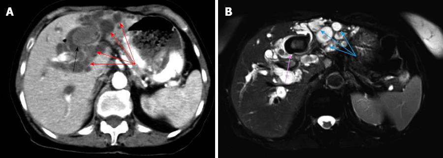
Figure 5 A case of Caroli disease.
A: On computed tomography. A large intra-biliary stone (black arrow) is evident in the dilated ducts (red arrows); B: On magnetic resonance imaging. A large intra-biliary stone (pink arrow) is evident in the dilated ducts (blue arrows).
- Citation: Bakoyiannis A, Delis S, Triantopoulou C, Dervenis C. Rare cystic liver lesions: A diagnostic and managing challenge. World J Gastroenterol 2013; 19(43): 7603-7619
- URL: https://www.wjgnet.com/1007-9327/full/v19/i43/7603.htm
- DOI: https://dx.doi.org/10.3748/wjg.v19.i43.7603


