Copyright
©2013 Baishideng Publishing Group Co.
World J Gastroenterol. Nov 21, 2013; 19(43): 7603-7619
Published online Nov 21, 2013. doi: 10.3748/wjg.v19.i43.7603
Published online Nov 21, 2013. doi: 10.3748/wjg.v19.i43.7603
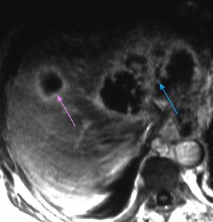
Figure 3 Magnetic resonance T1-w image shows an echinococcal cyst as a multiloculated cystic liver lesion, indicative of the presence of daughter cysts (light blue arrow).
A second smaller unilocular lesion with peripheral contrast enhancement is also seen (pink arrow).
- Citation: Bakoyiannis A, Delis S, Triantopoulou C, Dervenis C. Rare cystic liver lesions: A diagnostic and managing challenge. World J Gastroenterol 2013; 19(43): 7603-7619
- URL: https://www.wjgnet.com/1007-9327/full/v19/i43/7603.htm
- DOI: https://dx.doi.org/10.3748/wjg.v19.i43.7603


