Copyright
©2013 Baishideng Publishing Group Co.
World J Gastroenterol. Nov 14, 2013; 19(42): 7480-7486
Published online Nov 14, 2013. doi: 10.3748/wjg.v19.i42.7480
Published online Nov 14, 2013. doi: 10.3748/wjg.v19.i42.7480
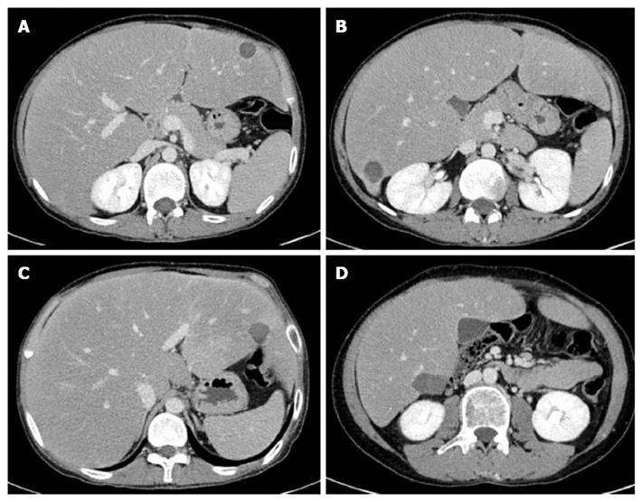
Figure 3 Computed tomogram follow-up after 3 mo showed gradual reduction of radiofrequency ablation zone.
A: Post-RFA follow-up CT revealed gradual contraction of ablated lesion in segment 3 to 1 cm; B: Complete ablation of enhancing lesion in segment 6 was observed in Post-RFA follow-up CT; C: Post-RFA follow-up CT showed complete ablation of 1.8 cm sized tumor in segment 2; D: Post-RFA CT revealed no residual lesion without adjacent organ damage. CT: Computed tomogram; RFA: Radiofrequency ablation.
- Citation: Ahn SY, Park SY, Kweon YO, Tak WY, Bae HI, Cho SH. Successful treatment of multiple hepatocellular adenomas with percutaneous radiofrequency ablation. World J Gastroenterol 2013; 19(42): 7480-7486
- URL: https://www.wjgnet.com/1007-9327/full/v19/i42/7480.htm
- DOI: https://dx.doi.org/10.3748/wjg.v19.i42.7480


