Copyright
©2013 Baishideng Publishing Group Co.
World J Gastroenterol. Nov 14, 2013; 19(42): 7480-7486
Published online Nov 14, 2013. doi: 10.3748/wjg.v19.i42.7480
Published online Nov 14, 2013. doi: 10.3748/wjg.v19.i42.7480
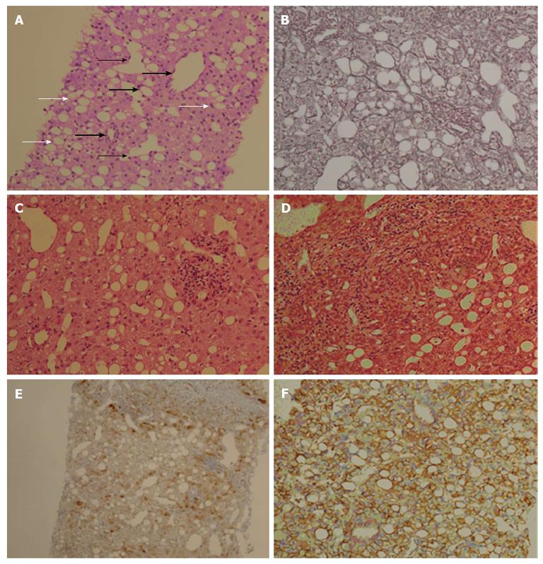
Figure 2 The histological features are consistent with the inflammatory type of hepatocellular adenoma.
A: A hepatocellular adenoma in GSD-Ia has several unpaired arteries (thick black arrows), sinusoidal dilation (thin black arrows), and steatosis (white arrows), but the atypia of hepatocytes are low without a nuclear pleomorphism; B: A reticulin staining does not show thick hepatic cords; C: The lymphocytic infiltrates are found in a hepatic lobule; D: The lymphocytes infiltrate in the area surrounded a portal and a periportal tracts; E: Staining for glutamine synthetase is diffuse cytoplasmic with focal nuclear expression; F: β-catenin staining does not show abnormal expression. GSD-Ia: Glycogen storage disease type Ia.
- Citation: Ahn SY, Park SY, Kweon YO, Tak WY, Bae HI, Cho SH. Successful treatment of multiple hepatocellular adenomas with percutaneous radiofrequency ablation. World J Gastroenterol 2013; 19(42): 7480-7486
- URL: https://www.wjgnet.com/1007-9327/full/v19/i42/7480.htm
- DOI: https://dx.doi.org/10.3748/wjg.v19.i42.7480


