Copyright
©2013 Baishideng Publishing Group Co.
World J Gastroenterol. Nov 14, 2013; 19(42): 7419-7425
Published online Nov 14, 2013. doi: 10.3748/wjg.v19.i42.7419
Published online Nov 14, 2013. doi: 10.3748/wjg.v19.i42.7419
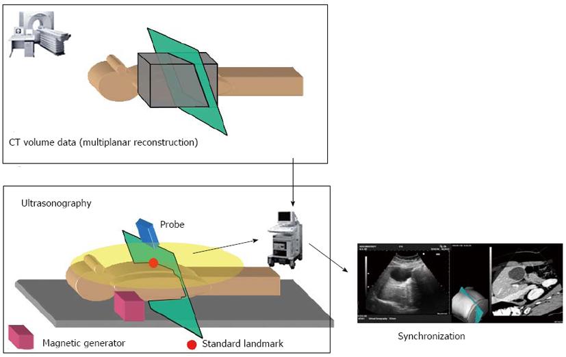
Figure 2 Calibration process.
Transfer of the obtained computed tomography (CT) volume data; configuration of the settings for the standard landmark (ensiform cartilage/aorta/portal vein/other); and positional information of the probe is sensed. According to the probe sensor, the multiplanar reconstruction image is shown as an optimal angled plane
- Citation: Sofuni A, Itoi T, Itokawa F, Tsuchiya T, Kurihara T, Ishii K, Tsuji S, Ikeuchi N, Tanaka R, Umeda J, Tonozuka R, Honjo M, Mukai S, Moriyasu F. Real-time virtual sonography visualization and its clinical application in biliopancreatic disease. World J Gastroenterol 2013; 19(42): 7419-7425
- URL: https://www.wjgnet.com/1007-9327/full/v19/i42/7419.htm
- DOI: https://dx.doi.org/10.3748/wjg.v19.i42.7419


