Copyright
©2013 Baishideng Publishing Group Co.
World J Gastroenterol. Nov 14, 2013; 19(42): 7389-7398
Published online Nov 14, 2013. doi: 10.3748/wjg.v19.i42.7389
Published online Nov 14, 2013. doi: 10.3748/wjg.v19.i42.7389
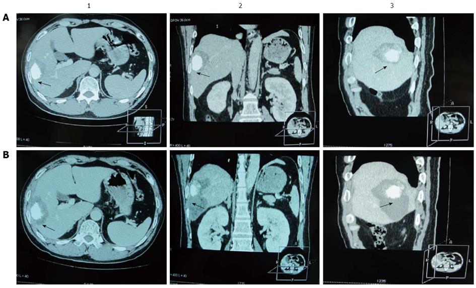
Figure 2 Three-dimensional computed tomography for radiofrequency ablation imaging in a patient with hepatocellular carcinoma.
A: The computed tomography (CT) images prior to the second radiofrequency (RF) ablation; B: The CT images after the second RF ablation. 1: Axial plane; 2: Coronal plane; 3: Sagittal plane. The ablative margin is < 0.5 cm prior to the second RF ablation, and it has been ablated to ≥ 0.5 cm after the second RF ablation. Black arrow indicates the shortest width of the area of low density outside the iodine stained tumor.
- Citation: Ke S, Ding XM, Qian XJ, Zhou YM, Cao BX, Gao K, Sun WB. Radiofrequency ablation of hepatocellular carcinoma sized > 3 and ≤ 5 cm: Is ablative margin of more than 1 cm justified? World J Gastroenterol 2013; 19(42): 7389-7398
- URL: https://www.wjgnet.com/1007-9327/full/v19/i42/7389.htm
- DOI: https://dx.doi.org/10.3748/wjg.v19.i42.7389


