Copyright
©2013 Baishideng Publishing Group Co.
World J Gastroenterol. Nov 14, 2013; 19(42): 7374-7388
Published online Nov 14, 2013. doi: 10.3748/wjg.v19.i42.7374
Published online Nov 14, 2013. doi: 10.3748/wjg.v19.i42.7374
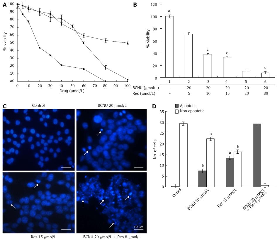
Figure 5 Anti-proliferative and apoptotic effect of 3-Bis(2-chloroethyl)-1-nitrosourea and/or resveratrol on 5-fluorouracil-R cells.
A: Anchorage-dependent cell survival of 5-FU-R cells after treatment with 5-FU, Res and BCNU; B: Bar diagram representing the % viability of 5-FU-R cells after BCNU + Res exposure. Data are the mean ± SD of three different experiments, aP < 0.05 vs 20 μmol/L BCNU + 5 μmol/L Res; cP < 0.05 vs 20 μmol/L BCNU + 20 μmol/L Res; C: Apoptotic nuclei after DAPI staining. Images were taken using a fluorescent microscope (Nikon-Eclipse, Japan) at ×40 magnification. An arrow indicates the apoptotic nuclei. Data are the representation of one of the replicates of three different experiments; D: A graphical representation of apoptotic nuclei, aP < 0.05 vs 20 μmol/L BCNU + 8 μmol/L Res. 5-FU: 5-fluorouracil; Res: Resveratrol; BCNU: 1,3-Bis(2-chloroethyl)-1-nitrosourea.
- Citation: Das D, Preet R, Mohapatra P, Satapathy SR, Kundu CN. 1,3-Bis(2-chloroethyl)-1-nitrosourea enhances the inhibitory effect of Resveratrol on 5-fluorouracil sensitive/resistant colon cancer cells. World J Gastroenterol 2013; 19(42): 7374-7388
- URL: https://www.wjgnet.com/1007-9327/full/v19/i42/7374.htm
- DOI: https://dx.doi.org/10.3748/wjg.v19.i42.7374


