Copyright
©2013 Baishideng Publishing Group Co.
World J Gastroenterol. Nov 14, 2013; 19(42): 7341-7360
Published online Nov 14, 2013. doi: 10.3748/wjg.v19.i42.7341
Published online Nov 14, 2013. doi: 10.3748/wjg.v19.i42.7341
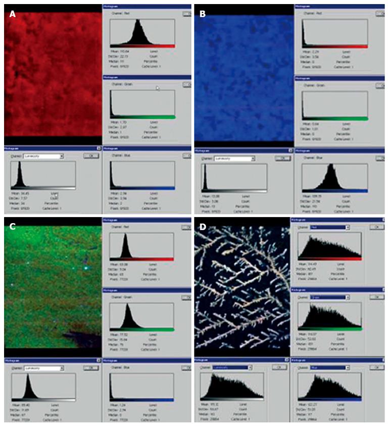
Figure 2 Color cathode-luminescence scanning electron microscopy images.
Color cathode-luminescence scanning electron microscopy (CCL SEM) micro images of unconjugated bilirubin (A), unesterified cholesterol (B), high-molecular-weight protein (C), and dehydrated sample of normal human cystic bile (D).
- Citation: Reshetnyak VI. Physiological and molecular biochemical mechanisms of bile formation. World J Gastroenterol 2013; 19(42): 7341-7360
- URL: https://www.wjgnet.com/1007-9327/full/v19/i42/7341.htm
- DOI: https://dx.doi.org/10.3748/wjg.v19.i42.7341


