Copyright
©2013 Baishideng Publishing Group Co.
World J Gastroenterol. Nov 14, 2013; 19(42): 7247-7257
Published online Nov 14, 2013. doi: 10.3748/wjg.v19.i42.7247
Published online Nov 14, 2013. doi: 10.3748/wjg.v19.i42.7247
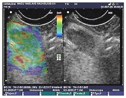
Figure 6 Endoscopic ultrasonography B-mode sonogram and elastogram of lymph node in chronic pancreatitis.
The sonogram (right) shows a lymph node as a hypoechoic oval shape surrounded by more echogenic tissue in the liver hilum. This lymph node approximately 18 mm × 10 mm, appeared in the liver hilum of a patient with chronic pancreatitis. On the left, the lymph node is not harder than the surrounding tissue as the predominant color hue is green. This finding is frequent in reactive lymph nodes, and may be a sign of benign etiology.
- Citation: Dimcevski G, Erchinger FG, Havre R, Gilja OH. Ultrasonography in diagnosing chronic pancreatitis: New aspects. World J Gastroenterol 2013; 19(42): 7247-7257
- URL: https://www.wjgnet.com/1007-9327/full/v19/i42/7247.htm
- DOI: https://dx.doi.org/10.3748/wjg.v19.i42.7247


