Copyright
©2013 Baishideng Publishing Group Co.
World J Gastroenterol. Nov 14, 2013; 19(42): 7247-7257
Published online Nov 14, 2013. doi: 10.3748/wjg.v19.i42.7247
Published online Nov 14, 2013. doi: 10.3748/wjg.v19.i42.7247
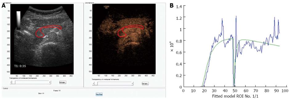
Figure 3 Perfusion analysis of the pancreas.
A: Dual view of contrast-enhanced ultrasonography examination of the pancreas in a healthy volunteer. 1.5 mL bolus of Sonovue was given as a bolus and after approximately 45 s the area of interest was exposed to high MI ultrasound bursting the bubbles in the imaging plane; B: A motion correcting analyzing software was used (DCE-US, http://www.isibrno.cz/perfusion/). A region of interest have been drawn including the head and body of the pancreas (unpublished data).
- Citation: Dimcevski G, Erchinger FG, Havre R, Gilja OH. Ultrasonography in diagnosing chronic pancreatitis: New aspects. World J Gastroenterol 2013; 19(42): 7247-7257
- URL: https://www.wjgnet.com/1007-9327/full/v19/i42/7247.htm
- DOI: https://dx.doi.org/10.3748/wjg.v19.i42.7247


