Copyright
©2013 Baishideng Publishing Group Co.
World J Gastroenterol. Nov 14, 2013; 19(42): 7247-7257
Published online Nov 14, 2013. doi: 10.3748/wjg.v19.i42.7247
Published online Nov 14, 2013. doi: 10.3748/wjg.v19.i42.7247
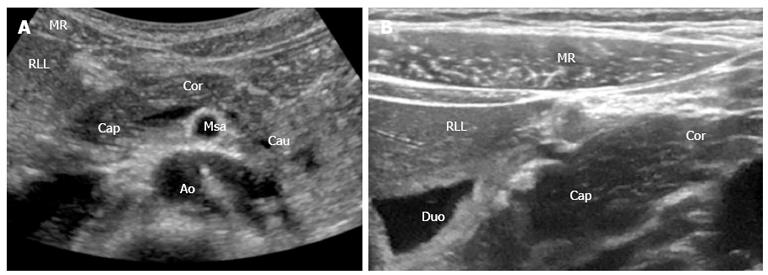
Figure 1 Pancreas and the surrounding anatomical landmarks.
A: B-mode image (1-5 MHz); B: B-mode image with a 12-15 MHz transducer. Details shown with high resolution. MR: Musculus rectus abdominis; RLL: Right liver lobe; Cap: Caput pancreatis; Cor: Corpus pancreatis; Cau: Cauda pancreatis; Msa: Superior mesenteric artery; Duo: Duodenum; Ao: Aorta.
- Citation: Dimcevski G, Erchinger FG, Havre R, Gilja OH. Ultrasonography in diagnosing chronic pancreatitis: New aspects. World J Gastroenterol 2013; 19(42): 7247-7257
- URL: https://www.wjgnet.com/1007-9327/full/v19/i42/7247.htm
- DOI: https://dx.doi.org/10.3748/wjg.v19.i42.7247


